4JLT | pdb_00004jlt

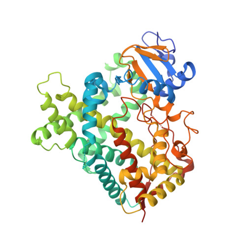
Crystal structure of P450 2B4(H226Y) in complex with paroxetine


Experimental Data Snapshot

  • Method: X-RAY DIFFRACTION
  • Resolution: 2.14 Å
  • R-Value Free: 
    0.229 (Depositor), 0.236 (DCC) 
  • R-Value Work: 
    0.187 (Depositor), 0.209 (DCC) 
  • R-Value Observed: 
    0.190 (Depositor) 

Starting Model: experimental
View more details

wwPDB Validation 3D Report Full Report

Validation slider image for 4JLT

Ligand Structure Quality Assessment 


This is version 1.3 of the entry. See complete history. 

Literature

A Structural Snapshot of CYP2B4 in Complex with Paroxetine Provides Insights into Ligand Binding and Clusters of Conformational States.

Shah, M.B., Kufareva, I., Pascual, J., Zhang, Q., Stout, C.D., Halpert, J.R.

(2013) J Pharmacol Exp Ther 346: 113-120

  • DOI: https://doi.org/10.1124/jpet.113.204776
  • Primary Citation Related Structures: 
    4JLT

  • PubMed Abstract: 

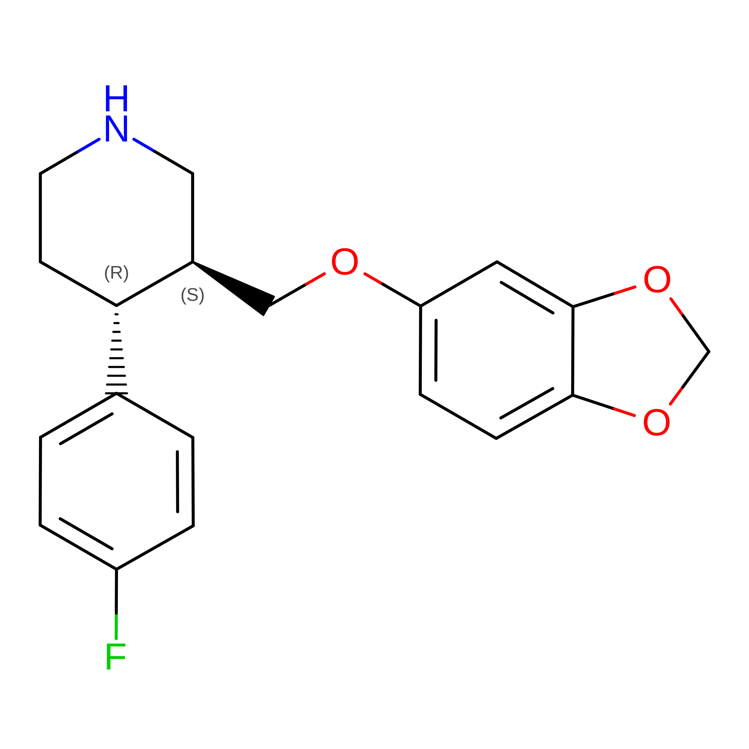
    An X-ray crystal structure of CYP2B4 in complex with the drug paroxetine [(3S,4R)-3-[(2H-1,3-benzodioxol-5-yloxy)methyl]-4-(4-fluorophenyl)piperidine] was solved at 2.14 Å resolution. The structure revealed a conformation intermediate to that of the recently solved complex with amlodipine and that of the more compact complex with 4-(4-chlorophenyl)imidazole in terms of the placement of the F-G cassette. Moreover, comparison of the new structure with 15 previously solved structures of CYP2B4 revealed some new insights into the determinants of active-site size and shape. The 2B4-paroxetine structure is nearly superimposable on a previously solved closed structure in a ligand-free state. Despite the overall conformational similarity among multiple closed structures, the active-site cavity volume of the paroxetine complex is enlarged. Further analysis of the accessible space and binding pocket near the heme reveals a new subchamber that resulted from the movement of secondary structural elements and rearrangements of active-site side chains. Overall, the results from the comparison of all 16 structures of CYP2B4 demonstrate a cluster of protein conformations that were observed in the presence or absence of various ligands.


  • Organizational Affiliation: 
    • Skaggs School of Pharmacy and Pharmaceutical Sciences, University of California San Diego, 9500 Gilman Drive, Mail Code 0703, La Jolla, CA 92093-0703, USA. m7shah@ucsd.edu

Macromolecule Content 

  • Total Structure Weight: 57.26 kDa 
  • Atom Count: 4,056 
  • Modeled Residue Count: 458 
  • Deposited Residue Count: 476 
  • Unique protein chains: 1

Macromolecules

Find similar proteins by:|  3D Structure
Entity ID: 1
MoleculeChains  Sequence LengthOrganismDetailsImage
Cytochrome P450 2B4476Oryctolagus cuniculusMutation(s): 9 
Gene Names: CYP2B4
EC: 1.14.14.1
UniProt
Find proteins for P00178 (Oryctolagus cuniculus)
Explore P00178 
Go to UniProtKB:  P00178
Entity Groups
Sequence Clusters30% Identity50% Identity70% Identity90% Identity95% Identity100% Identity
UniProt GroupP00178
Sequence Annotations
Expand
Reference Sequence

Small Molecules

Ligands 6 Unique
IDChains Name / Formula / InChI Key2D Diagram3D Interactions
HEM

Query on HEM


Download:Ideal Coordinates CCD File
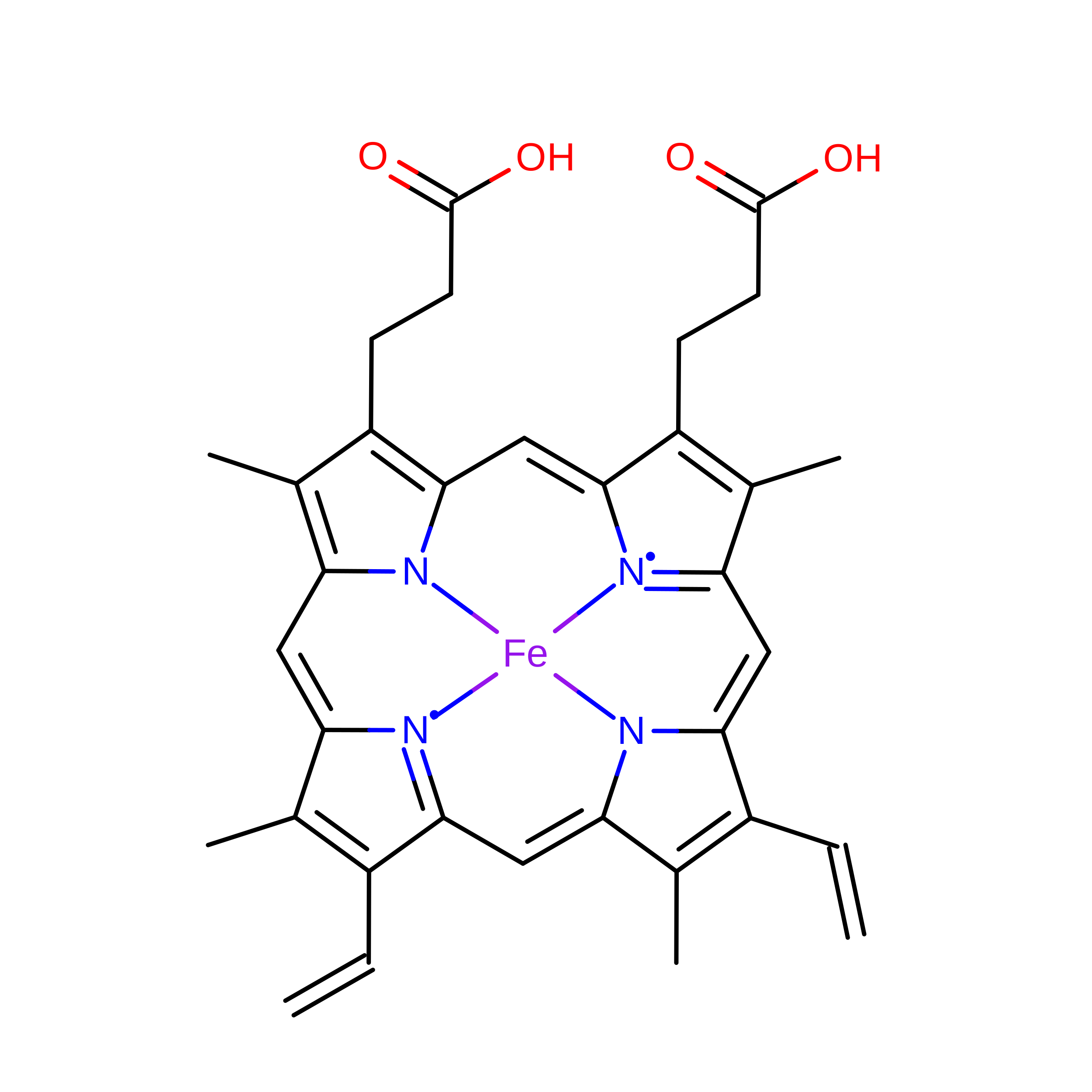
B [auth A]PROTOPORPHYRIN IX CONTAINING FE
C34 H32 Fe N4 O4
KABFMIBPWCXCRK-RGGAHWMASA-L
CM5

Query on CM5


Download:Ideal Coordinates CCD File
C [auth A],
D [auth A],
E [auth A]
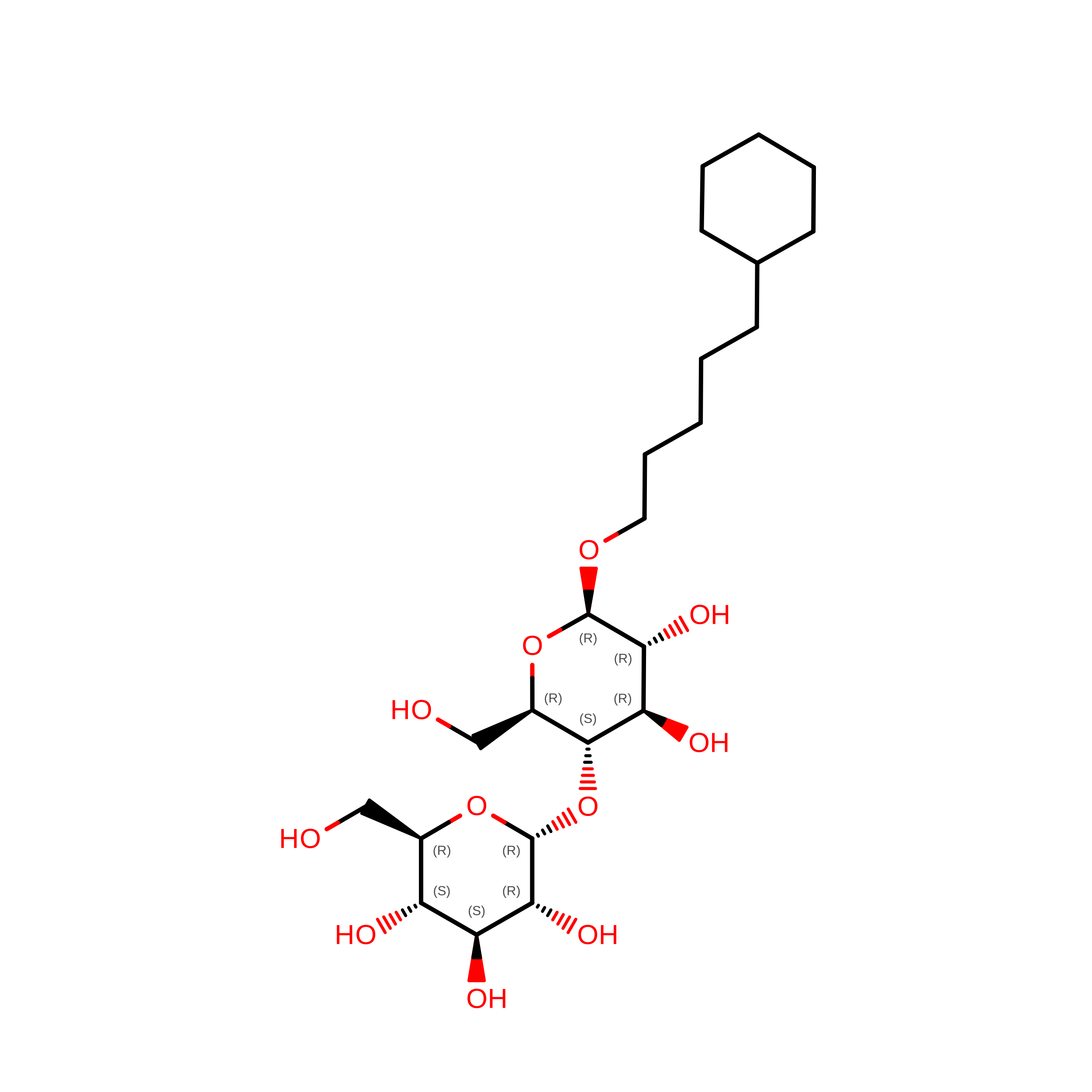
5-CYCLOHEXYL-1-PENTYL-BETA-D-MALTOSIDE
C23 H42 O11
RVTGFZGNOSKUDA-ZNGNCRBCSA-N
8PR

Query on 8PR


Download:Ideal Coordinates CCD File
F [auth A]Paroxetine
C19 H20 F N O3
AHOUBRCZNHFOSL-YOEHRIQHSA-N
SO4

Query on SO4


Download:Ideal Coordinates CCD File
G [auth A],
H [auth A],
I [auth A],
J [auth A]
SULFATE ION
O4 S
QAOWNCQODCNURD-UHFFFAOYSA-L
PO4

Query on PO4


Download:Ideal Coordinates CCD File
L [auth A],
M [auth A]
PHOSPHATE ION
O4 P
NBIIXXVUZAFLBC-UHFFFAOYSA-K
GOL

Query on GOL


Download:Ideal Coordinates CCD File
K [auth A]GLYCEROL
C3 H8 O3
PEDCQBHIVMGVHV-UHFFFAOYSA-N

Experimental Data & Validation

Experimental Data

  • Method: X-RAY DIFFRACTION
  • Resolution: 2.14 Å
  • R-Value Free:  0.229 (Depositor), 0.236 (DCC) 
  • R-Value Work:  0.187 (Depositor), 0.209 (DCC) 
  • R-Value Observed: 0.190 (Depositor) 
Space Group: P 31 2 1
Unit Cell:
Length ( Å )Angle ( ˚ )
a = 90.76α = 90
b = 90.76β = 90
c = 153.681γ = 120
Software Package:
Software NamePurpose
REFMACrefinement
PDB_EXTRACTdata extraction
Blu-Icedata collection
iMOSFLMdata reduction
SCALAdata scaling
PHASERphasing

Structure Validation

View Full Validation Report


Ligand Structure Quality Assessment 


Entry History 

Deposition Data

Revision History  (Full details and data files)

  • Version 1.0: 2013-06-26
    Type: Initial release
  • Version 1.1: 2013-07-03
    Changes: Database references
  • Version 1.2: 2023-09-20
    Changes: Data collection, Database references, Derived calculations, Refinement description
  • Version 1.3: 2026-08-12
    Type: Remediation
    Reason: Metalloprotein remediation
    Changes: Derived calculations, Structure summary