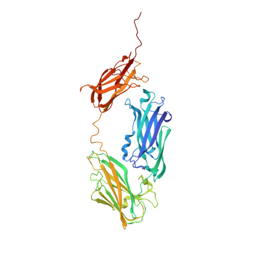
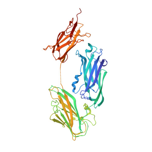
Structures of SdrD from Staphylococcus aureus reveal the molecular mechanism of how the cell surface receptors recognize their ligands
Wang, X., Ge, J., Liu, B., Hu, Y., Yang, M.(2013) Protein Cell 4: 277-285
- PubMed: 23549613 Search on PubMedSearch on PubMed Central
- DOI: https://doi.org/10.1007/s13238-013-3009-x
- Primary Citation Related Structures:
4JDZ, 4JE0 - PubMed Abstract:
Staphylococcus aureus is the most important Gram-positive colonizer of human skin and nasal passage, causing high morbidity and mortality. SD-repeat containing protein D (SdrD), an MSCRAMM (Microbial Surface Components Recognizing Adhesive Matrix Molecules) family surface protein, plays an important role in S. aureus adhesion and pathogenesis, while its binding target and molecular mechanism remain largely unknown. Here we solved the crystal structures of SdrD N2-N3 domain and N2-N3-B1 domain. Through structural analysis and comparisons, we characterized the ligand binding site of SdrD, and proposed a featured sequence motif of its potential ligands. In addition, the structures revealed for the first time the interactions between B1 domain and N2-N3 domain among B domain-containing MSCRAMMs. Our results may help in understanding the roles SdrD plays in S. aureus adhesion and shed light on the development of novel antibiotics.
- Key Laboratory for Protein Sciences of Ministry of Education, Tsinghua-Peking Center for Life Sciences, School of Life Sciences, Tsinghua University, Beijing 100084, China.
Organizational Affiliation: