4JBW

Crystal structure of E. coli maltose transporter MalFGK2 in complex with its regulatory protein EIIAglc


Experimental Data Snapshot

  • Method: X-RAY DIFFRACTION
  • Resolution: 3.91 Å
  • R-Value Free: 0.281 
  • R-Value Work: 0.226 
  • R-Value Observed: 0.229 

wwPDB Validation   3D Report Full Report


Ligand Structure Quality Assessment 


This is version 1.2 of the entry. See complete history


Literature

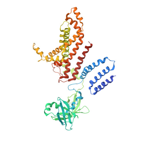
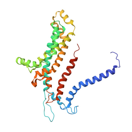
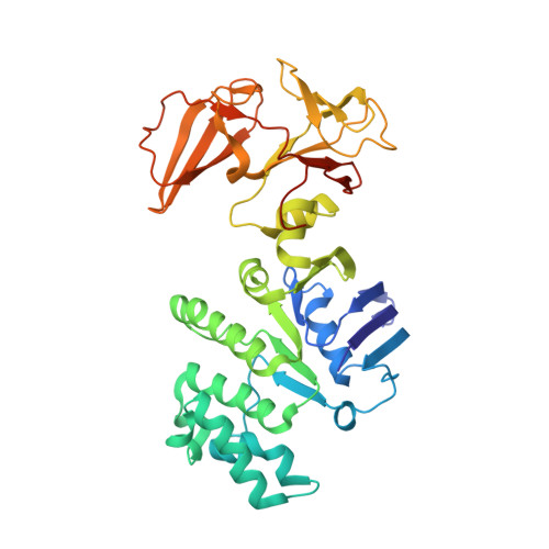
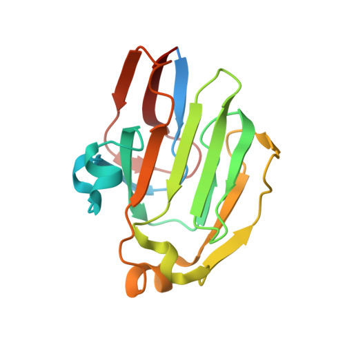
Carbon catabolite repression of the maltose transporter revealed by X-ray crystallography.

Chen, S.Oldham, M.L.Davidson, A.L.Chen, J.

(2013) Nature 499: 364-368

  • DOI: https://doi.org/10.1038/nature12232
  • Primary Citation of Related Structures:  
    4JBW

  • PubMed Abstract: 

    Efficient carbon utilization is critical to the survival of microorganisms in competitive environments. To optimize energy usage, bacteria have developed an integrated control system to preferentially uptake carbohydrates that support rapid growth. The availability of a preferred carbon source, such as glucose, represses the synthesis and activities of proteins necessary for the transport and metabolism of secondary carbon sources. This regulatory phenomenon is defined as carbon catabolite repression. In enteric bacteria, the key player of carbon catabolite repression is a component of the glucose-specific phosphotransferase system, enzyme IIA (EIIA(Glc)). It is known that unphosphorylated EIIA(Glc) binds to and inhibits a variety of transporters when glucose is available. However, understanding the underlying molecular mechanism has been hindered by the complete absence of structures for any EIIA(Glc)-transporter complexes. Here we present the 3.9 Å crystal structure of Escherichia coli EIIA(Glc) in complex with the maltose transporter, an ATP-binding cassette (ABC) transporter. The structure shows that two EIIA(Glc) molecules bind to the cytoplasmic ATPase subunits, stabilizing the transporter in an inward-facing conformation and preventing the structural rearrangements necessary for ATP hydrolysis. We also show that the half-maximal inhibitory concentrations of the full-length EIIA(Glc) and an amino-terminal truncation mutant differ by 60-fold, consistent with the hypothesis that the amino-terminal region, disordered in the crystal structure, functions as a membrane anchor to increase the effective EIIA(Glc) concentration at the membrane. Together these data suggest a model of how the central regulatory protein EIIA(Glc) allosterically inhibits maltose uptake in E. coli.


  • Organizational Affiliation

    Department of Biological Sciences, Purdue University, West Lafayette, Indiana 47907, USA.


Macromolecules
Find similar proteins by:  (by identity cutoff)  |  3D Structure
Entity ID: 1
MoleculeChains Sequence LengthOrganismDetailsImage
Maltose transport system permease protein MalFA [auth F],
G [auth H]
514Escherichia coli K-12Mutation(s): 0 
Gene Names: malFb4033JW3993
Membrane Entity: Yes 
UniProt
Find proteins for P02916 (Escherichia coli (strain K12))
Explore P02916 
Go to UniProtKB:  P02916
Entity Groups  
Sequence Clusters30% Identity50% Identity70% Identity90% Identity95% Identity100% Identity
UniProt GroupP02916
Sequence Annotations
Expand
  • Reference Sequence
Find similar proteins by:  (by identity cutoff)  |  3D Structure
Entity ID: 2
MoleculeChains Sequence LengthOrganismDetailsImage
Maltose transport system permease protein MalGB [auth G],
H [auth I]
296Escherichia coli K-12Mutation(s): 0 
Gene Names: malGb4032JW3992
Membrane Entity: Yes 
UniProt
Find proteins for P68183 (Escherichia coli (strain K12))
Explore P68183 
Go to UniProtKB:  P68183
Entity Groups  
Sequence Clusters30% Identity50% Identity70% Identity90% Identity95% Identity100% Identity
UniProt GroupP68183
Sequence Annotations
Expand
  • Reference Sequence
Find similar proteins by:  (by identity cutoff)  |  3D Structure
Entity ID: 3
MoleculeChains Sequence LengthOrganismDetailsImage
Maltose/maltodextrin import ATP-binding protein MalKC [auth A],
D [auth B],
I [auth C],
J [auth D]
381Escherichia coli K-12Mutation(s): 0 
Gene Names: malKb4035JW3995
EC: 3.6.3.19
Membrane Entity: Yes 
UniProt
Find proteins for P68187 (Escherichia coli (strain K12))
Explore P68187 
Go to UniProtKB:  P68187
Entity Groups  
Sequence Clusters30% Identity50% Identity70% Identity90% Identity95% Identity100% Identity
UniProt GroupP68187
Sequence Annotations
Expand
  • Reference Sequence
Find similar proteins by:  (by identity cutoff)  |  3D Structure
Entity ID: 4
MoleculeChains Sequence LengthOrganismDetailsImage
Glucose-specific phosphotransferase enzyme IIA componentE [auth M],
F [auth N],
K [auth O],
L [auth P]
172Escherichia coli K-12Mutation(s): 0 
Gene Names: crrgsriextgstreDb2417JW2410
EC: 2.7.1
UniProt
Find proteins for P69783 (Escherichia coli (strain K12))
Explore P69783 
Go to UniProtKB:  P69783
Entity Groups  
Sequence Clusters30% Identity50% Identity70% Identity90% Identity95% Identity100% Identity
UniProt GroupP69783
Sequence Annotations
Expand
  • Reference Sequence
Small Molecules
Ligands 1 Unique
IDChains Name / Formula / InChI Key2D Diagram3D Interactions
PGV
Query on PGV

Download Ideal Coordinates CCD File 
M [auth F],
N [auth H]
(1R)-2-{[{[(2S)-2,3-DIHYDROXYPROPYL]OXY}(HYDROXY)PHOSPHORYL]OXY}-1-[(PALMITOYLOXY)METHYL]ETHYL (11E)-OCTADEC-11-ENOATE
C40 H77 O10 P
ADYWCMPUNIVOEA-GPJPVTGXSA-N
Experimental Data & Validation

Experimental Data

  • Method: X-RAY DIFFRACTION
  • Resolution: 3.91 Å
  • R-Value Free: 0.281 
  • R-Value Work: 0.226 
  • R-Value Observed: 0.229 
  • Space Group: P 21 21 21
Unit Cell:
Length ( Å )Angle ( ˚ )
a = 94.529α = 90
b = 208.448β = 90
c = 347.477γ = 90
Software Package:
Software NamePurpose
HKL-2000data collection
PHASERphasing
PHENIXrefinement
HKL-2000data reduction
HKL-2000data scaling

Structure Validation

View Full Validation Report


Ligand Structure Quality Assessment 


Entry History 

Deposition Data

Revision History  (Full details and data files)

  • Version 1.0: 2013-06-12
    Type: Initial release
  • Version 1.1: 2013-07-17
    Changes: Database references
  • Version 1.2: 2023-09-20
    Changes: Data collection, Database references, Derived calculations, Refinement description, Structure summary