4JA0

Crystal structure of the invertebrate bi-functional purine biosynthesis enzyme PAICS at 2.8 A resolution


Experimental Data Snapshot

  • Method: X-RAY DIFFRACTION
  • Resolution: 2.80 Å
  • R-Value Free: 0.263 
  • R-Value Work: 0.199 
  • R-Value Observed: 0.203 

wwPDB Validation   3D Report Full Report


This is version 1.3 of the entry. See complete history


Literature

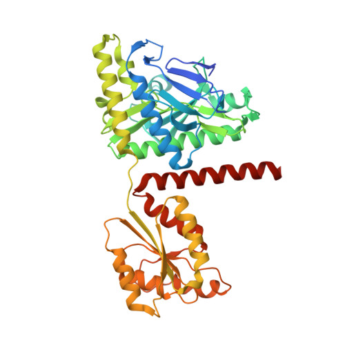
Crystal structure of the invertebrate bifunctional purine biosynthesis enzyme PAICS at 2.8 angstrom resolution.

Taschner, M.Basquin, J.Benda, C.Lorentzen, E.

(2013) Proteins 81: 1473-1478

  • DOI: https://doi.org/10.1002/prot.24296
  • Primary Citation of Related Structures:  
    4JA0

  • PubMed Abstract: 

    Two important steps of the de novo purine biosynthesis pathway are catalyzed by the 5-aminoimidazole ribonucleotide carboxylase and the 4-(N-succinylcarboxamide)-5-aminoimidazole ribonucleotide synthetase enzymes. In most eukaryotic organisms, these two activities are present in the bifunctional enzyme complex known as PAICS. We have determined the 2.8-Å resolution crystal structure of the 350-kDa invertebrate PAICS from insect cells (Trichoplusia ni) using single-wavelength anomalous dispersion methods. Comparison of insect PAICS to human and prokaryotic homologs provides insights into substrate binding and reveals a highly conserved enzymatic framework across divergent species.


  • Organizational Affiliation

    Max-Planck-Institute of Biochemistry, Department of Structural Cell Biology, Martinsried, Germany.


Macromolecules
Find similar proteins by:  (by identity cutoff)  |  3D Structure
Entity ID: 1
MoleculeChains Sequence LengthOrganismDetailsImage
Phosphoribosylaminoimidazole carboxylase
A, B, C, D
425Bombyx moriMutation(s): 0 
UniProt
Find proteins for Q1HQ66 (Bombyx mori)
Explore Q1HQ66 
Go to UniProtKB:  Q1HQ66
Entity Groups  
Sequence Clusters30% Identity50% Identity70% Identity90% Identity95% Identity100% Identity
UniProt GroupQ1HQ66
Sequence Annotations
Expand
  • Reference Sequence
Small Molecules
Ligands 1 Unique
IDChains Name / Formula / InChI Key2D Diagram3D Interactions
SO4
Query on SO4

Download Ideal Coordinates CCD File 
E [auth A]
F [auth A]
G [auth A]
H [auth A]
I [auth A]
E [auth A],
F [auth A],
G [auth A],
H [auth A],
I [auth A],
J [auth A],
K [auth B],
L [auth B],
M [auth B],
N [auth B],
O [auth C],
P [auth C],
Q [auth C],
R [auth C],
S [auth D],
T [auth D],
U [auth D]
SULFATE ION
O4 S
QAOWNCQODCNURD-UHFFFAOYSA-L
Experimental Data & Validation

Experimental Data

  • Method: X-RAY DIFFRACTION
  • Resolution: 2.80 Å
  • R-Value Free: 0.263 
  • R-Value Work: 0.199 
  • R-Value Observed: 0.203 
  • Space Group: P 21 21 2
Unit Cell:
Length ( Å )Angle ( ˚ )
a = 138.729α = 90
b = 219.726β = 90
c = 60.645γ = 90
Software Package:
Software NamePurpose
PHENIXrefinement

Structure Validation

View Full Validation Report


Entry History 

Deposition Data

Revision History  (Full details and data files)

  • Version 1.0: 2013-02-27
    Type: Initial release
  • Version 1.1: 2013-04-10
    Changes: Database references
  • Version 1.2: 2013-10-23
    Changes: Database references
  • Version 1.3: 2023-09-20
    Changes: Data collection, Database references, Derived calculations, Refinement description