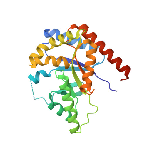
Structural and biochemical properties of LuxF from Photobacterium leiognathi.
Bergner, T., Tabib, C.R., Winkler, A., Stipsits, S., Kayer, H., Lee, J., Malthouse, J.P., Mayhew, S., Muller, F., Gruber, K., Macheroux, P.(2015) Biochim Biophys Acta 1854: 1466-1475
- PubMed: 26209460 Search on PubMed
- DOI: https://doi.org/10.1016/j.bbapap.2015.07.008
- Primary Citation Related Structures:
4J2P - PubMed Abstract:
The lux-operon of bioluminescent bacteria contains the genes coding for the enzymes required for light emission. Some species of Photobacteria feature an additional gene, luxF, which shows similarity to luxA and luxB, the genes encoding the heterodimeric luciferase. Isolated dimeric LuxF binds four molecules of an unusually derivatized flavin, i.e., 6-(3'-(R)-myristyl)-FMN (myrFMN). In the present study we have heterologously expressed LuxF in Escherichia coli BL21 in order to advance our understanding of the protein's binding properties and its role in photobacterial bioluminescence. Structure determination by X-ray crystallography confirmed that apo-LuxF possesses four preorganized binding sites, which are further optimized by adjusting the orientation of amino acid side chains. To investigate the binding properties of recombinant LuxF we have isolated myrFMN from Photobacterium leiognathi S1. We found that LuxF binds myrFMN tightly with a dissociation constant of 80±20 nM demonstrating that the purified apo-form of LuxF is fully competent in myrFMN binding. In contrast to LuxF, binding of myrFMN to luciferase is much weaker (Kd=4.0±0.4 μM) enabling LuxF to prevent inhibition of the enzyme by scavenging myrFMN. Moreover, we have used apo-LuxF to demonstrate that myrFMN occurs in all Photobacteria tested, irrespective of the presence of luxF indicating that LuxF is not required for myrFMN biosynthesis.
- Graz University of Technology, Institute of Biochemistry, Graz, Austria.
Organizational Affiliation: