


Crystal structure analysis of Thermus thermophilus transhydrogenase soluble domains
Yamaguchi, M., Leung, J., Schurig Briccio, L.A., Gennis, R.B., Stout, C.D.To be published.
Experimental Data Snapshot
Starting Models: experimental
View more details
Entity ID: 1 | |||||
|---|---|---|---|---|---|
| Molecule | Chains | Sequence Length | Organism | Details | Image |
| NAD/NADP transhydrogenase alpha subunit 1 | 381 | Thermus thermophilus HB27 | Mutation(s): 0 Gene Names: TT_C1780 EC: 1.6.1.2 (PDB Primary Data), 7.1.1.1 (UniProt) | 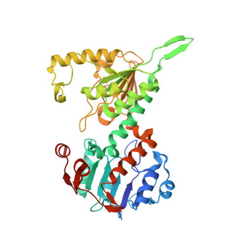 | |
UniProt | |||||
Entity Groups | |||||
| Sequence Clusters | 30% Identity50% Identity70% Identity90% Identity95% Identity100% Identity | ||||
| UniProt Group | Q72GR8 | ||||
Sequence AnnotationsExpand | |||||
Reference Sequence | |||||
Entity ID: 2 | |||||
|---|---|---|---|---|---|
| Molecule | Chains | Sequence Length | Organism | Details | Image |
| NAD(P) transhydrogenase subunit beta | 185 | Thermus thermophilus HB27 | Mutation(s): 0 EC: 1.6.1.2 (PDB Primary Data), 7.1.1.1 (UniProt) | 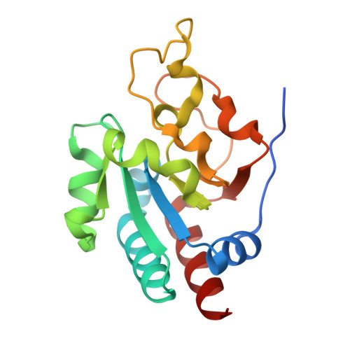 | |
UniProt | |||||
Entity Groups | |||||
| Sequence Clusters | 30% Identity50% Identity70% Identity90% Identity95% Identity100% Identity | ||||
| UniProt Group | Q72GS0 | ||||
Sequence AnnotationsExpand | |||||
Reference Sequence | |||||
| Ligands 5 Unique | |||||
|---|---|---|---|---|---|
| ID | Chains | Name / Formula / InChI Key | 2D Diagram | 3D Interactions | |
| NAP Download:Ideal Coordinates CCD File | I [auth C] | NADP NICOTINAMIDE-ADENINE-DINUCLEOTIDE PHOSPHATE C21 H28 N7 O17 P3 XJLXINKUBYWONI-NNYOXOHSSA-N |  | ||
| NAD Download:Ideal Coordinates CCD File | D [auth A], E [auth B] | NICOTINAMIDE-ADENINE-DINUCLEOTIDE C21 H27 N7 O14 P2 BAWFJGJZGIEFAR-NNYOXOHSSA-N |  | ||
| PGE Download:Ideal Coordinates CCD File | G [auth B] | TRIETHYLENE GLYCOL C6 H14 O4 ZIBGPFATKBEMQZ-UHFFFAOYSA-N |  | ||
| GOL Download:Ideal Coordinates CCD File | H [auth B], J [auth C] | GLYCEROL C3 H8 O3 PEDCQBHIVMGVHV-UHFFFAOYSA-N |  | ||
| CL Download:Ideal Coordinates CCD File | F [auth B] | CHLORIDE ION Cl VEXZGXHMUGYJMC-UHFFFAOYSA-M |  | ||
| Length ( Å ) | Angle ( ˚ ) |
|---|---|
| a = 67.616 | α = 90 |
| b = 75.038 | β = 90 |
| c = 198.144 | γ = 90 |
| Software Name | Purpose |
|---|---|
| ADSC | data collection |
| PHASER | phasing |
| REFMAC | refinement |
| XDS | data reduction |
| SCALA | data scaling |


