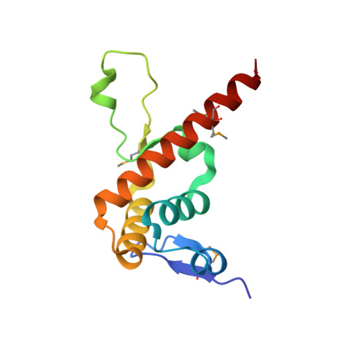
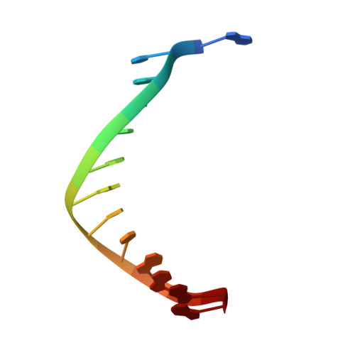
The BEN domain is a novel sequence-specific DNA-binding domain conserved in neural transcriptional repressors.
Dai, Q., Ren, A., Westholm, J.O., Serganov, A.A., Patel, D.J., Lai, E.C.(2013) Genes Dev 27: 602-614
- PubMed: 23468431 Search on PubMedSearch on PubMed Central
- DOI: https://doi.org/10.1101/gad.213314.113
- Primary Citation Related Structures:
4IX7 - PubMed Abstract:
We recently reported that Drosophila Insensitive (Insv) promotes sensory organ development and has activity as a nuclear corepressor for the Notch transcription factor Suppressor of Hairless [Su(H)]. Insv lacks domains of known biochemical function but contains a single BEN domain (i.e., a "BEN-solo" protein). Our chromatin immunoprecipitation (ChIP) sequencing (ChIP-seq) analysis confirmed binding of Insensitive to Su(H) target genes in the Enhancer of split gene complex [E(spl)-C]; however, de novo motif analysis revealed a novel site strongly enriched in Insv peaks (TCYAATHRGAA). We validate binding of endogenous Insv to genomic regions bearing such sites, whose associated genes are enriched for neural functions and are functionally repressed by Insv. Unexpectedly, we found that the Insv BEN domain binds specifically to this sequence motif and that Insv directly regulates transcription via this motif. We determined the crystal structure of the BEN-DNA target complex, revealing homodimeric binding of the BEN domain and extensive nucleotide contacts via α helices and a C-terminal loop. Point mutations in key DNA-contacting residues severely impair DNA binding in vitro and capacity for transcriptional regulation in vivo. We further demonstrate DNA-binding and repression activities by the mammalian neural BEN-solo protein BEND5. Altogether, we define novel DNA-binding activity in a conserved family of transcriptional repressors, opening a molecular window on this extensive gene family.
- Department of Developmental Biology, Sloan-Kettering Institute, New York, New York 10065, USA.
Organizational Affiliation: