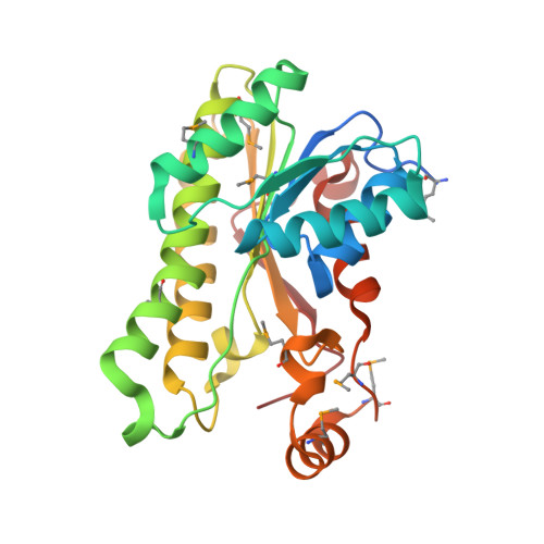
Crystal structure of a 3alpha-hydroxysteroid dehydrogenase (BaiA2) associated with secondary bile acid synthesis from Clostridium scindens VPI12708 in complex with a putative NAD(+)-OH- adduct at 2.0 A resolution
Joint Center for Structural Genomics (JCSG)To be published.