4IP9 | pdb_00004ip9

Structure of native human serum amyloid A1


Experimental Data Snapshot

  • Method: X-RAY DIFFRACTION
  • Resolution: 2.50 Å
  • R-Value Free: 
    0.258 (Depositor), 0.281 (DCC) 
  • R-Value Work: 
    0.249 (Depositor), 0.256 (DCC) 
  • R-Value Observed: 
    0.250 (Depositor) 

wwPDB Validation 3D Report Full Report

Validation slider image for 4IP9

Ligand Structure Quality Assessment 


This is version 1.3 of the entry. See complete history

Literature

Structural mechanism of serum amyloid A-mediated inflammatory amyloidosis.

Lu, J.Yu, Y.Zhu, I.Cheng, Y.Sun, P.D.

(2014) Proc Natl Acad Sci U S A 111: 5189-5194

  • DOI: https://doi.org/10.1073/pnas.1322357111
  • Primary Citation Related Structures: 
    4IP8, 4IP9

  • PubMed Abstract: 

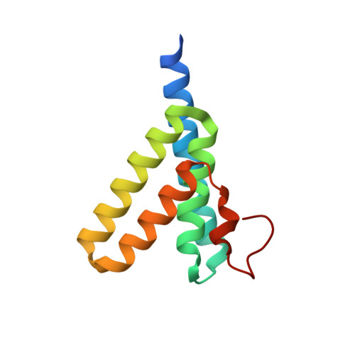
    Serum amyloid A (SAA) represents an evolutionarily conserved family of inflammatory acute-phase proteins. It is also a major constituent of secondary amyloidosis. To understand its function and structural transition to amyloid, we determined a structure of human SAA1.1 in two crystal forms, representing a prototypic member of the family. Native SAA1.1 exists as a hexamer, with subunits displaying a unique four-helix bundle fold stabilized by its long C-terminal tail. Structure-based mutational studies revealed two positive-charge clusters, near the center and apex of the hexamer, that are involved in SAA association with heparin. The binding of high-density lipoprotein involves only the apex region of SAA and can be inhibited by heparin. Peptide amyloid formation assays identified the N-terminal helices 1 and 3 as amyloidogenic peptides of SAA1.1. Both peptides are secluded in the hexameric structure of SAA1.1, suggesting that the native SAA is nonpathogenic. Furthermore, dissociation of the SAA hexamer appears insufficient to initiate amyloidogenic transition, and proteolytic cleavage or removal of the C-terminal tail of SAA resulted in formation of various-sized structural aggregates containing ∼5-nm regular repeating protofibril-like units. The combined structural and functional studies provide mechanistic insights into the pathogenic contribution of glycosaminoglycan in SAA1.1-mediated AA amyloid formation.


  • Organizational Affiliation
    • Structural Immunology Section, Laboratory of Immunogenetics, National Institute of Allergy and Infectious Diseases, National Institutes of Health, Rockville, MD 20852.

Macromolecule Content 

  • Total Structure Weight: 27.27 kDa 
  • Atom Count: 1,744 
  • Modeled Residue Count: 208 
  • Deposited Residue Count: 222 
  • Unique protein chains: 1

Macromolecules

Find similar proteins by:|  3D Structure
Entity ID: 1
MoleculeChains  Sequence LengthOrganismDetailsImage
Serum amyloid A-1 protein
A, B
111Homo sapiensMutation(s): 0 
Gene Names: SAA1
UniProt & NIH Common Fund Data Resources
Find proteins for P0DJI8 (Homo sapiens)
Explore P0DJI8 
Go to UniProtKB:  P0DJI8
PHAROS:  P0DJI8
GTEx:  ENSG00000173432 
Entity Groups
Sequence Clusters30% Identity50% Identity70% Identity90% Identity95% Identity100% Identity
UniProt GroupP0DJI8
Sequence Annotations
Expand
Reference Sequence

Experimental Data & Validation

Experimental Data

  • Method: X-RAY DIFFRACTION
  • Resolution: 2.50 Å
  • R-Value Free:  0.258 (Depositor), 0.281 (DCC) 
  • R-Value Work:  0.249 (Depositor), 0.256 (DCC) 
  • R-Value Observed: 0.250 (Depositor) 
Space Group: H 3 2
Unit Cell:
Length ( Å )Angle ( ˚ )
a = 93.85α = 90
b = 93.85β = 90
c = 130.52γ = 120
Software Package:
Software NamePurpose
HKL-2000data collection
PHASERphasing
BUSTERrefinement
HKL-2000data reduction
HKL-2000data scaling

Structure Validation

View Full Validation Report


Ligand Structure Quality Assessment 


Entry History 

Deposition Data

Revision History  (Full details and data files)

  • Version 1.0: 2014-03-26
    Type: Initial release
  • Version 1.1: 2014-05-14
    Changes: Database references
  • Version 1.2: 2018-01-24
    Changes: Structure summary
  • Version 1.3: 2024-02-28
    Changes: Data collection, Database references, Derived calculations