4IP1 | pdb_00004ip1

C-terminal domain of the thiol:disulfide interchange protein DsbD, Q488K mutant


Experimental Data Snapshot

  • Method: X-RAY DIFFRACTION
  • Resolution: 2.47 Å
  • R-Value Free: 
    0.255 (Depositor), 0.248 (DCC) 
  • R-Value Work: 
    0.193 (Depositor), 0.194 (DCC) 
  • R-Value Observed: 
    0.196 (Depositor) 

Starting Model: experimental
View more details

wwPDB Validation 3D Report Full Report

Validation slider image for 4IP1

This is version 1.5 of the entry. See complete history

Literature

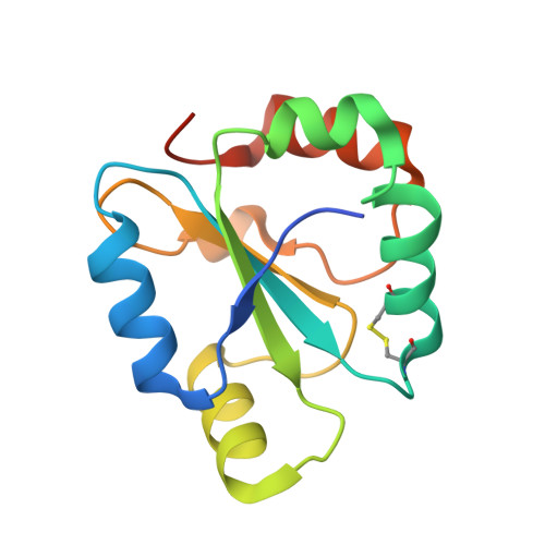
An Extended Active-site Motif Controls the Reactivity of the Thioredoxin Fold.

Mavridou, D.A.Saridakis, E.Kritsiligkou, P.Mozley, E.C.Ferguson, S.J.Redfield, C.

(2014) J Biological Chem 289: 8681-8696

  • DOI: https://doi.org/10.1074/jbc.M113.513457
  • Primary Citation Related Structures: 
    4IP1, 4IP6

  • PubMed Abstract: 

    Proteins belonging to the thioredoxin (Trx) superfamily are abundant in all organisms. They share the same structural features, arranged in a seemingly simple fold, but they perform a multitude of functions in oxidative protein folding and electron transfer pathways. We use the C-terminal domain of the unique transmembrane reductant conductor DsbD as a model for an in-depth analysis of the factors controlling the reactivity of the Trx fold. We employ NMR spectroscopy, x-ray crystallography, mutagenesis, in vivo functional experiments applied to DsbD, and a comparative sequence analysis of Trx-fold proteins to determine the effect of residues in the vicinity of the active site on the ionization of the key nucleophilic cysteine of the -CXXC- motif. We show that the function and reactivity of Trx-fold proteins depend critically on the electrostatic features imposed by an extended active-site motif.


  • Organizational Affiliation
    • From the Department of Biochemistry, University of Oxford, South Parks Road, Oxford OX1 3QU, United Kingdom and.

Macromolecule Content 

  • Total Structure Weight: 14.63 kDa 
  • Atom Count: 1,062 
  • Modeled Residue Count: 121 
  • Deposited Residue Count: 132 
  • Unique protein chains: 1

Macromolecules

Find similar proteins by:|  3D Structure
Entity ID: 1
MoleculeChains  Sequence LengthOrganismDetailsImage
Thiol:disulfide interchange protein DsbD132Escherichia coli K-12Mutation(s): 1 
Gene Names: dsbDcutA2cycZdipZb4136JW5734
EC: 1.8.1.8
UniProt
Find proteins for P36655 (Escherichia coli (strain K12))
Explore P36655 
Go to UniProtKB:  P36655
Entity Groups
Sequence Clusters30% Identity50% Identity70% Identity90% Identity95% Identity100% Identity
UniProt GroupP36655
Sequence Annotations
Expand
Reference Sequence

Experimental Data & Validation

Experimental Data

  • Method: X-RAY DIFFRACTION
  • Resolution: 2.47 Å
  • R-Value Free:  0.255 (Depositor), 0.248 (DCC) 
  • R-Value Work:  0.193 (Depositor), 0.194 (DCC) 
  • R-Value Observed: 0.196 (Depositor) 
Space Group: P 21 21 21
Unit Cell:
Length ( Å )Angle ( ˚ )
a = 35.107α = 90
b = 51.53β = 90
c = 85.039γ = 90
Software Package:
Software NamePurpose
MOLREPphasing
PHENIXrefinement
DENZOdata reduction
SCALEPACKdata scaling

Structure Validation

View Full Validation Report


Entry History 

Deposition Data

Revision History  (Full details and data files)

  • Version 1.0: 2013-12-25
    Type: Initial release
  • Version 1.1: 2014-02-12
    Changes: Database references
  • Version 1.2: 2014-04-09
    Changes: Database references
  • Version 1.3: 2017-11-15
    Changes: Advisory, Refinement description
  • Version 1.4: 2023-09-20
    Changes: Advisory, Data collection, Database references, Refinement description
  • Version 1.5: 2024-11-20
    Changes: Structure summary