4II2

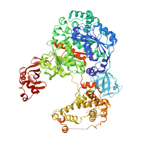
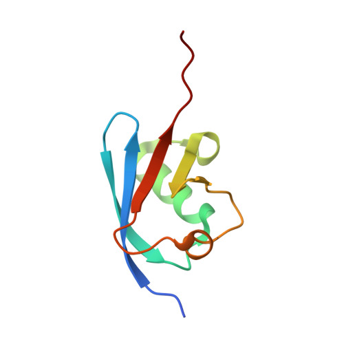
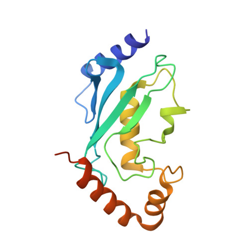
Crystal structure of Ubiquitin activating enzyme 1 (Uba1) in complex with the Ub E2 Ubc4, ubiquitin, and ATP/Mg


Experimental Data Snapshot

  • Method: X-RAY DIFFRACTION
  • Resolution: 2.20 Å
  • R-Value Free: 0.254 
  • R-Value Work: 0.213 
  • R-Value Observed: 0.213 

Starting Model: experimental
View more details

wwPDB Validation   3D Report Full Report


Ligand Structure Quality Assessment 


This is version 1.3 of the entry. See complete history


Literature

Structure of a ubiquitin E1-E2 complex: insights to E1-E2 thioester transfer.

Olsen, S.K.Lima, C.D.

(2013) Mol Cell 49: 884-896

  • DOI: https://doi.org/10.1016/j.molcel.2013.01.013
  • Primary Citation of Related Structures:  
    4II2, 4II3

  • PubMed Abstract: 

    Ubiquitin (Ub) conjugation is initiated by an E1 enzyme that catalyzes carboxy-terminal Ub adenylation, thioester bond formation to a catalytic cysteine in the E1 Cys domain, and thioester transfer to a catalytic cysteine in E2 conjugating enzymes. How the E1 and E2 active sites come together during thioester transfer and how Ub E1 interacts with diverse Ub E2s remains unclear. Here we present a crystal structure of a Ub E1-E2(Ubc4)/Ub/ATP·Mg complex that was stabilized by induction of a disulfide bond between the E1 and E2 active sites. The structure reveals combinatorial recognition of the E2 by the E1 ubiquitin-fold domain (UFD) and Cys domain and mutational analysis, coupled with thioester transfer assays with E1, Ubc4, and other Ub E2s, show that both interfaces are important for thioester transfer. Comparison to a Ub E1/Ub/ATP·Mg structure reveals conformational changes in the E1 that bring the E1 and E2 active sites together.


  • Organizational Affiliation

    Structural Biology Program, Sloan-Kettering Institute, New York, NY 10065, USA.


Macromolecules
Find similar proteins by:  (by identity cutoff)  |  3D Structure
Entity ID: 1
MoleculeChains Sequence LengthOrganismDetailsImage
Ubiquitin-activating enzyme E1 11,001Schizosaccharomyces pombe 972h-Mutation(s): 0 
Gene Names: ptr3SPBC1604.21cSPBC211.09Ubiquitin activating enzyme 1 (Uba1)
EC: 6.3.2.19
UniProt
Find proteins for O94609 (Schizosaccharomyces pombe (strain 972 / ATCC 24843))
Explore O94609 
Go to UniProtKB:  O94609
Entity Groups  
Sequence Clusters30% Identity50% Identity70% Identity90% Identity95% Identity100% Identity
UniProt GroupO94609
Sequence Annotations
Expand
  • Reference Sequence
Find similar proteins by:  (by identity cutoff)  |  3D Structure
Entity ID: 2
MoleculeChains Sequence LengthOrganismDetailsImage
Ubiquitin-60S ribosomal protein L4083Schizosaccharomyces pombe 972h-Mutation(s): 9 
Gene Names: ubi2
UniProt
Find proteins for P0CH07 (Schizosaccharomyces pombe (strain 972 / ATCC 24843))
Explore P0CH07 
Go to UniProtKB:  P0CH07
Entity Groups  
Sequence Clusters30% Identity50% Identity70% Identity90% Identity95% Identity100% Identity
UniProt GroupP0CH07
Sequence Annotations
Expand
  • Reference Sequence
Find similar proteins by:  (by identity cutoff)  |  3D Structure
Entity ID: 3
MoleculeChains Sequence LengthOrganismDetailsImage
Ubiquitin-conjugating enzyme E2 4163Schizosaccharomyces pombe 972h-Mutation(s): 2 
Gene Names: SPBC119.02ubc4Ubiquitin conjugating enzyme 4 (Ubc4)
EC: 6.3.2.19
UniProt
Find proteins for P46595 (Schizosaccharomyces pombe (strain 972 / ATCC 24843))
Explore P46595 
Go to UniProtKB:  P46595
Entity Groups  
Sequence Clusters30% Identity50% Identity70% Identity90% Identity95% Identity100% Identity
UniProt GroupP46595
Sequence Annotations
Expand
  • Reference Sequence
Small Molecules
Ligands 5 Unique
IDChains Name / Formula / InChI Key2D Diagram3D Interactions
ATP
Query on ATP

Download Ideal Coordinates CCD File 
F [auth A]ADENOSINE-5'-TRIPHOSPHATE
C10 H16 N5 O13 P3
ZKHQWZAMYRWXGA-KQYNXXCUSA-N
PG0
Query on PG0

Download Ideal Coordinates CCD File 
G [auth A]2-(2-METHOXYETHOXY)ETHANOL
C5 H12 O3
SBASXUCJHJRPEV-UHFFFAOYSA-N
SO4
Query on SO4

Download Ideal Coordinates CCD File 
N [auth A],
O [auth A],
P [auth A],
R [auth B]
SULFATE ION
O4 S
QAOWNCQODCNURD-UHFFFAOYSA-L
EDO
Query on EDO

Download Ideal Coordinates CCD File 
H [auth A]
I [auth A]
J [auth A]
K [auth A]
L [auth A]
H [auth A],
I [auth A],
J [auth A],
K [auth A],
L [auth A],
M [auth A],
Q [auth B]
1,2-ETHANEDIOL
C2 H6 O2
LYCAIKOWRPUZTN-UHFFFAOYSA-N
MG
Query on MG

Download Ideal Coordinates CCD File 
D [auth A],
E [auth A]
MAGNESIUM ION
Mg
JLVVSXFLKOJNIY-UHFFFAOYSA-N
Experimental Data & Validation

Experimental Data

  • Method: X-RAY DIFFRACTION
  • Resolution: 2.20 Å
  • R-Value Free: 0.254 
  • R-Value Work: 0.213 
  • R-Value Observed: 0.213 
  • Space Group: P 21 21 21
Unit Cell:
Length ( Å )Angle ( ˚ )
a = 81.6α = 90
b = 111.2β = 90
c = 181.3γ = 90
Software Package:
Software NamePurpose
DENZOdata reduction
SCALEPACKdata scaling
CNSrefinement
PDB_EXTRACTdata extraction
HKL-2000data collection
PHASERphasing

Structure Validation

View Full Validation Report


Ligand Structure Quality Assessment 


Entry History 

Deposition Data

Revision History  (Full details and data files)

  • Version 1.0: 2013-02-13
    Type: Initial release
  • Version 1.1: 2013-03-27
    Changes: Database references
  • Version 1.2: 2017-11-15
    Changes: Advisory, Refinement description
  • Version 1.3: 2023-09-20
    Changes: Advisory, Data collection, Database references, Derived calculations, Refinement description