4IF7

Mycobacterium Tuberculosis Methionine aminopeptidase Type 1c in complex with homocysteine-methyl disulfide


Experimental Data Snapshot

  • Method: X-RAY DIFFRACTION
  • Resolution: 2.00 Å
  • R-Value Free: 0.266 
  • R-Value Work: 0.201 
  • R-Value Observed: 0.205 

wwPDB Validation   3D Report Full Report


Ligand Structure Quality Assessment 


This is version 1.3 of the entry. See complete history


Literature

Selective targeting of the conserved active site cysteine of Mycobacterium tuberculosis methionine aminopeptidase with electrophilic reagents

Reddi, R.Arya, T.Kishor, C.Gumpena, R.Ganji, R.J.Bhukya, S.Addlagatta, A.

(2014) FEBS J 281: 4240-4248

  • DOI: https://doi.org/10.1111/febs.12847
  • Primary Citation of Related Structures:  
    4IDY, 4IEC, 4IF7, 4OOK

  • PubMed Abstract: 

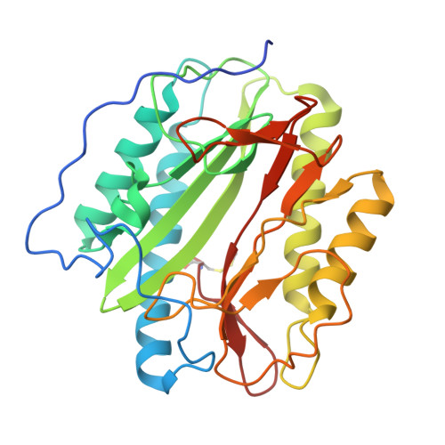
    Methionine aminopeptidases (MetAPs) cleave initiator methionine from ~ 70% of the newly synthesized proteins in every living cell, and specific inhibition or knockdown of this function is detrimental. MetAPs are metalloenzymes, and are broadly classified into two subtypes, type I and type II. Bacteria contain only type I MetAPs, and the active site of these enzymes contains a conserved cysteine. By contrast, in type II enzymes the analogous position is occupied by a conserved glycine. Here, we report the reactivity of the active site cysteine in a type I MetAP, MetAP1c, of Mycobacterium tuberculosis (MtMetAP1c) towards highly selective cysteine-specific reagents. The authenticity of selective modification of Cys105 of MtMetAP1c was established by using site-directed mutagenesis and crystal structure determination of covalent and noncovalent complexes. On the basis of these observations, we propose that metal ions in the active site assist in the covalent modification of Cys105 by orienting the reagents appropriately for a successful reaction. These studies establish, for the first time, that the conserved cysteine of type I MetAPs can be targeted for selective inhibition, and we believe that this chemistry can be exploited for further drug discovery efforts regarding microbial MetAPs.


  • Organizational Affiliation

    Center for Chemical Biology, CSIR-Indian Institute of Chemical Technology, Hyderabad, Telengana, India.


Macromolecules
Find similar proteins by:  (by identity cutoff)  |  3D Structure
Entity ID: 1
MoleculeChains Sequence LengthOrganismDetailsImage
Methionine aminopeptidase 2291Mycobacterium tuberculosisMutation(s): 0 
Gene Names: map
EC: 3.4.11.18
UniProt
Find proteins for P9WK19 (Mycobacterium tuberculosis (strain ATCC 25618 / H37Rv))
Explore P9WK19 
Go to UniProtKB:  P9WK19
Entity Groups  
Sequence Clusters30% Identity50% Identity70% Identity90% Identity95% Identity100% Identity
UniProt GroupP9WK19
Sequence Annotations
Expand
  • Reference Sequence
Small Molecules
Modified Residues  1 Unique
IDChains TypeFormula2D DiagramParent
SCH
Query on SCH
A
L-PEPTIDE LINKINGC4 H9 N O2 S2CYS
Experimental Data & Validation

Experimental Data

  • Method: X-RAY DIFFRACTION
  • Resolution: 2.00 Å
  • R-Value Free: 0.266 
  • R-Value Work: 0.201 
  • R-Value Observed: 0.205 
  • Space Group: P 1 21 1
Unit Cell:
Length ( Å )Angle ( ˚ )
a = 49.38α = 90
b = 48.216β = 95.25
c = 56.4γ = 90
Software Package:
Software NamePurpose
HKL-3000data collection
AMoREphasing
REFMACrefinement
DENZOdata reduction
SCALEPACKdata scaling

Structure Validation

View Full Validation Report


Ligand Structure Quality Assessment 


Entry History 

Deposition Data

Revision History  (Full details and data files)

  • Version 1.0: 2013-12-18
    Type: Initial release
  • Version 1.1: 2014-04-16
    Changes: Other
  • Version 1.2: 2015-02-04
    Changes: Database references
  • Version 1.3: 2019-11-20
    Changes: Database references, Derived calculations