


Crystal Structure of Staphylococcal nuclease mutant V23I/I72L
Hansen, S.W., Janowska, K., Stites, W.E., Sakon, J.To be published.
Experimental Data Snapshot
Starting Model: experimental
View more details
wwPDB Validation 3D Report Full Report

Entity ID: 1 | |||||
|---|---|---|---|---|---|
| Molecule | Chains | Sequence Length | Organism | Details | Image |
| Thermonuclease | 149 | Staphylococcus aureus | Mutation(s): 2 Gene Names: nuc EC: 3.1.31.1 | 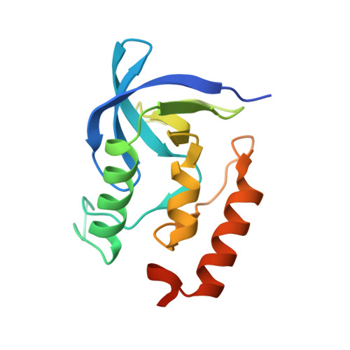 | |
UniProt | |||||
Entity Groups | |||||
| Sequence Clusters | 30% Identity50% Identity70% Identity90% Identity95% Identity100% Identity | ||||
| UniProt Group | P00644 | ||||
Sequence AnnotationsExpand | |||||
Reference Sequence | |||||
| Length ( Å ) | Angle ( ˚ ) |
|---|---|
| a = 48.3 | α = 90 |
| b = 48.3 | β = 90 |
| c = 63.9 | γ = 90 |
| Software Name | Purpose |
|---|---|
| CrystalClear | data collection |
| CCP4 | model building |
| REFMAC | refinement |
| d*TREK | data reduction |
| d*TREK | data scaling |
| CCP4 | phasing |


