4ICY

Tracing the Evolution of Angucyclinone Monooxygenases: Structural Determinants for C-12b Hydroxylation and Substrate Inhibition in PgaE


Experimental Data Snapshot

  • Method: X-RAY DIFFRACTION
  • Resolution: 2.40 Å
  • R-Value Free: 0.259 
  • R-Value Work: 0.198 
  • R-Value Observed: 0.202 

wwPDB Validation   3D Report Full Report


Ligand Structure Quality Assessment 


This is version 1.3 of the entry. See complete history


Literature

Tracing the Evolution of Angucyclinone Monooxygenases: Structural Determinants for C-12b Hydroxylation and Substrate Inhibition in PgaE.

Kallio, P.Patrikainen, P.Belogurov, G.A.Mantsala, P.Yang, K.Niemi, J.Metsa-Ketela, M.

(2013) Biochemistry 52: 4507-4516

  • DOI: https://doi.org/10.1021/bi400381s
  • Primary Citation of Related Structures:  
    4ICY

  • PubMed Abstract: 

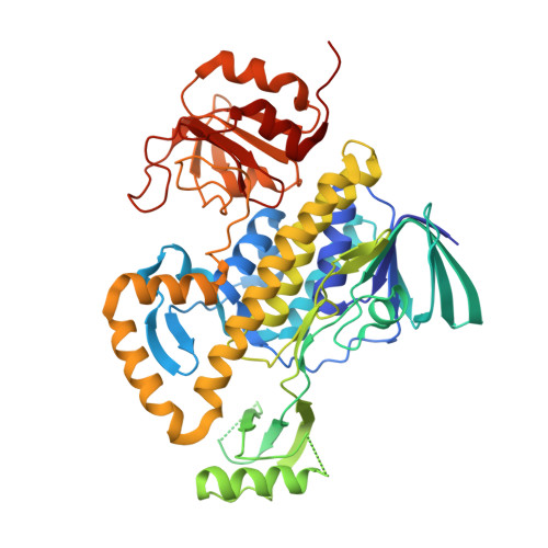
    Two functionally distinct homologous flavoprotein hydroxylases, PgaE and JadH, have been identified as branching points in the biosynthesis of the polyketide antibiotics gaudimycin C and jadomycin A, respectively. These evolutionarily related enzymes are both bifunctional and able to catalyze the same initial reaction, C-12 hydroxylation of the common angucyclinone intermediate prejadomycin. The enzymes diverge in their secondary activities, which include hydroxylation at C-12b by PgaE and dehydration at C-4a/C-12b by JadH. A further difference is that the C-12 hydroxylation is subject to substrate inhibition only in PgaE. Here we have identified regions associated with the C-12b hydroxylation in PgaE by extensive chimeragenesis, focusing on regions surrounding the active site. The results highlight the importance of a hairpin-β motif near the dimer interface, with two nonconserved residues, P78 and I79 (corresponding to Q89 and F90, respectively, in JadH), and invariant residue H73 playing key roles. Kinetic characterization of PgaE variants demonstrates that the secondary C-12b hydroxylation and substrate inhibition by prejadomycin are likely to be interlinked. The crystal structure of the PgaE P78Q/I79F variant at 2.4 Å resolution confirms that the changes do not alter the conformation of the β-strand secondary structure and that the side chains of these residues in effect point away from the active site toward the dimer interface. The results support a catalytic model for PgaE containing two binding modes for C-12 and C-12b hydroxylations, where binding of prejadomycin in the orientation for C-12b hydroxylation leads to substrate inhibition. The presence of an allosteric network is evident based on enzyme kinetics.


  • Organizational Affiliation

    Department of Biochemistry and Food Chemistry, University of Turku, FIN-20014 Turku, Finland.


Macromolecules
Find similar proteins by:  (by identity cutoff)  |  3D Structure
Entity ID: 1
MoleculeChains Sequence LengthOrganismDetailsImage
Polyketide oxygenase PgaE500Streptomyces sp. PGA64Mutation(s): 2 
Gene Names: pgaE
UniProt
Find proteins for Q93LY7 (Streptomyces sp. PGA64)
Explore Q93LY7 
Go to UniProtKB:  Q93LY7
Entity Groups  
Sequence Clusters30% Identity50% Identity70% Identity90% Identity95% Identity100% Identity
UniProt GroupQ93LY7
Sequence Annotations
Expand
  • Reference Sequence
Small Molecules
Ligands 5 Unique
IDChains Name / Formula / InChI Key2D Diagram3D Interactions
FAD
Query on FAD

Download Ideal Coordinates CCD File 
B [auth A]FLAVIN-ADENINE DINUCLEOTIDE
C27 H33 N9 O15 P2
VWWQXMAJTJZDQX-UYBVJOGSSA-N
SO4
Query on SO4

Download Ideal Coordinates CCD File 
H [auth A],
I [auth A]
SULFATE ION
O4 S
QAOWNCQODCNURD-UHFFFAOYSA-L
GOL
Query on GOL

Download Ideal Coordinates CCD File 
E [auth A],
F [auth A]
GLYCEROL
C3 H8 O3
PEDCQBHIVMGVHV-UHFFFAOYSA-N
EDO
Query on EDO

Download Ideal Coordinates CCD File 
C [auth A],
D [auth A],
G [auth A],
J [auth A]
1,2-ETHANEDIOL
C2 H6 O2
LYCAIKOWRPUZTN-UHFFFAOYSA-N
CL
Query on CL

Download Ideal Coordinates CCD File 
K [auth A],
L [auth A]
CHLORIDE ION
Cl
VEXZGXHMUGYJMC-UHFFFAOYSA-M
Experimental Data & Validation

Experimental Data

  • Method: X-RAY DIFFRACTION
  • Resolution: 2.40 Å
  • R-Value Free: 0.259 
  • R-Value Work: 0.198 
  • R-Value Observed: 0.202 
  • Space Group: F 2 2 2
Unit Cell:
Length ( Å )Angle ( ˚ )
a = 65.97α = 90
b = 171.04β = 90
c = 212.88γ = 90
Software Package:
Software NamePurpose
EDNAdata collection
MOLREPphasing
REFMACrefinement
MOSFLMdata reduction
SCALAdata scaling

Structure Validation

View Full Validation Report


Ligand Structure Quality Assessment 


Entry History 

Deposition Data

Revision History  (Full details and data files)

  • Version 1.0: 2013-06-12
    Type: Initial release
  • Version 1.1: 2013-07-10
    Changes: Database references
  • Version 1.2: 2024-02-28
    Changes: Data collection, Database references, Derived calculations
  • Version 1.3: 2024-04-03
    Changes: Refinement description