4I9I

Crystal structure of tankyrase 1 with compound 4


Experimental Data Snapshot

  • Method: X-RAY DIFFRACTION
  • Resolution: 2.40 Å
  • R-Value Free: 0.286 
  • R-Value Work: 0.250 

wwPDB Validation   3D Report Full Report


Ligand Structure Quality Assessment 


This is version 1.1 of the entry. See complete history


Literature

Discovery of a class of novel tankyrase inhibitors that bind to both the nicotinamide pocket and the induced pocket.

Bregman, H.Gunaydin, H.Gu, Y.Schneider, S.Wilson, C.DiMauro, E.F.Huang, X.

(2013) J Med Chem 56: 1341-1345

  • DOI: https://doi.org/10.1021/jm301607v
  • Primary Citation of Related Structures:  
    4I9I

  • PubMed Abstract: 

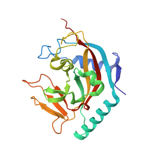
    Potent and selective inhibitors of tankyrases have recently been characterized to bind to an induced pocket. Here we report the identification of a novel potent and selective tankyrase inhibitor that binds to both the nicotinamide pocket and the induced pocket. The crystal structure of human TNKS1 in complex with this "dual-binder" provides a molecular basis for their strong and specific interactions and suggests clues for the further development of tankyrase inhibitors.


  • Organizational Affiliation

    Department of Medicinal Chemistry, Amgen Inc., 360 Binney Street, Cambridge, Massachusetts 02142, USA.


Macromolecules
Find similar proteins by:  (by identity cutoff)  |  3D Structure
Entity ID: 1
MoleculeChains Sequence LengthOrganismDetailsImage
Tankyrase-1
A, B, C, D
217Homo sapiensMutation(s): 0 
Gene Names: TNKSPARP5APARPLTIN1TINF1TNKS1
EC: 2.4.2.30
UniProt & NIH Common Fund Data Resources
Find proteins for O95271 (Homo sapiens)
Explore O95271 
Go to UniProtKB:  O95271
PHAROS:  O95271
GTEx:  ENSG00000173273 
Entity Groups  
Sequence Clusters30% Identity50% Identity70% Identity90% Identity95% Identity100% Identity
UniProt GroupO95271
Sequence Annotations
Expand
  • Reference Sequence
Small Molecules
Binding Affinity Annotations 
IDSourceBinding Affinity
1DY PDBBind:  4I9I IC50: 8 (nM) from 1 assay(s)
BindingDB:  4I9I IC50: min: 2, max: 8 (nM) from 2 assay(s)
Binding MOAD:  4I9I IC50: 8 (nM) from 1 assay(s)
Experimental Data & Validation

Experimental Data

  • Method: X-RAY DIFFRACTION
  • Resolution: 2.40 Å
  • R-Value Free: 0.286 
  • R-Value Work: 0.250 
  • Space Group: P 21 21 2
Unit Cell:
Length ( Å )Angle ( ˚ )
a = 158.622α = 90
b = 77.122β = 90
c = 84.507γ = 90
Software Package:
Software NamePurpose
HKL-2000data collection
AMoREphasing
CNSrefinement
HKL-2000data reduction
HKL-2000data scaling

Structure Validation

View Full Validation Report


Ligand Structure Quality Assessment 


Entry History 

Deposition Data

  • Released Date: 2013-02-06 
  • Deposition Author(s): Huang, X.

Revision History  (Full details and data files)

  • Version 1.0: 2013-02-06
    Type: Initial release
  • Version 1.1: 2013-05-22
    Changes: Database references