4I79

Crystal structure of human NUP43


Experimental Data Snapshot

  • Method: X-RAY DIFFRACTION
  • Resolution: 1.75 Å
  • R-Value Free: 0.212 
  • R-Value Work: 0.181 
  • R-Value Observed: 0.182 

wwPDB Validation   3D Report Full Report


This is version 1.5 of the entry. See complete history


Literature

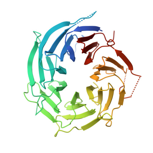
Crystal structure of human nuclear pore complex component NUP43.

Xu, C.Li, Z.He, H.Seitova, A.Wernimont, A.Li, Y.Loppnau, P.Min, J.

(2015) FEBS Lett 589: 3247-3253

  • DOI: https://doi.org/10.1016/j.febslet.2015.09.008

  • PubMed Abstract: 

    Nuclear pore complexes (NPC) form nuclear pores that cross the nuclear envelope and allow molecules to transport between the nucleus and the cytoplasm. We solved the crystal structure of human Nup43 (hNUP43), an important component in the Nup107 subcomplex of NPC. hNup43 adopts a seven-bladed β-propeller fold. We confirmed by ITC that neither human Nup37 (hNup37) nor human Nup133 (hNup133) interacts with hNup43. We demonstrated by analytical gel filtration that the human Nup85-Seh1L binary complex recruits hNup43 to form a ternary complex. Based on amino acid sequence analysis, we predicted the hNup85-hSeh1L binding surface of hNup43.


  • Organizational Affiliation

    Structural Genomics Consortium, University of Toronto, 101 College St., Toronto, Ontario M5G 1L7, Canada; Hefei National Laboratory for Physical Sciences at Microscale and School of Life Sciences, University of Science and Technology of China, Hefei, Anhui 230027, People's Republic of China.


Macromolecules
Find similar proteins by:  (by identity cutoff)  |  3D Structure
Entity ID: 1
MoleculeChains Sequence LengthOrganismDetailsImage
Nucleoporin Nup43
A, B
399Homo sapiensMutation(s): 0 
Gene Names: NUP43
UniProt & NIH Common Fund Data Resources
Find proteins for Q8NFH3 (Homo sapiens)
Explore Q8NFH3 
Go to UniProtKB:  Q8NFH3
PHAROS:  Q8NFH3
GTEx:  ENSG00000120253 
Entity Groups  
Sequence Clusters30% Identity50% Identity70% Identity90% Identity95% Identity100% Identity
UniProt GroupQ8NFH3
Sequence Annotations
Expand
  • Reference Sequence

Find similar proteins by:  Sequence   |   3D Structure  

Entity ID: 2
MoleculeChains Sequence LengthOrganismDetailsImage
floating chain, unknown sequence9Homo sapiensMutation(s): 0 
Sequence Annotations
Expand
  • Reference Sequence
Small Molecules
Ligands 1 Unique
IDChains Name / Formula / InChI Key2D Diagram3D Interactions
UNX
Query on UNX

Download Ideal Coordinates CCD File 
AA [auth B]
BA [auth B]
CA [auth B]
D [auth A]
DA [auth B]
AA [auth B],
BA [auth B],
CA [auth B],
D [auth A],
DA [auth B],
E [auth A],
EA [auth B],
F [auth A],
FA [auth B],
G [auth A],
GA [auth B],
H [auth A],
HA [auth B],
I [auth A],
IA [auth B],
J [auth A],
K [auth A],
L [auth A],
M [auth A],
N [auth A],
O [auth A],
P [auth A],
Q [auth B],
R [auth B],
S [auth B],
T [auth B],
U [auth B],
V [auth B],
W [auth B],
X [auth B],
Y [auth B],
Z [auth B]
UNKNOWN ATOM OR ION
X
Experimental Data & Validation

Experimental Data

  • Method: X-RAY DIFFRACTION
  • Resolution: 1.75 Å
  • R-Value Free: 0.212 
  • R-Value Work: 0.181 
  • R-Value Observed: 0.182 
  • Space Group: P 21 21 2
Unit Cell:
Length ( Å )Angle ( ˚ )
a = 80.736α = 90
b = 161.964β = 90
c = 58.197γ = 90
Software Package:
Software NamePurpose
SCALAdata scaling
PHASERphasing
REFMACrefinement
PDB_EXTRACTdata extraction
XDSdata reduction

Structure Validation

View Full Validation Report


Entry History 

Deposition Data

Revision History  (Full details and data files)

  • Version 1.0: 2013-02-27
    Type: Initial release
  • Version 1.1: 2013-11-27
    Changes: Other
  • Version 1.2: 2015-10-14
    Changes: Database references
  • Version 1.3: 2015-12-30
    Changes: Database references
  • Version 1.4: 2017-05-10
    Changes: Other, Structure summary
  • Version 1.5: 2023-09-20
    Changes: Data collection, Database references, Refinement description