4I60 | pdb_00004i60

Crystal structure of avidin - biotinylruthenocene complex


Experimental Data Snapshot

  • Method: X-RAY DIFFRACTION
  • Resolution: 2.50 Å
  • R-Value Free: 
    0.211 (Depositor), 0.212 (DCC) 
  • R-Value Work: 
    0.165 (Depositor), 0.166 (DCC) 
  • R-Value Observed: 
    0.167 (Depositor) 

Starting Model: experimental
View more details

wwPDB Validation 3D Report Full Report

Validation slider image for 4I60

Ligand Structure Quality Assessment 


This is version 2.2 of the entry. See complete history

Literature

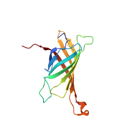
Structural investigation of the interactions of biotinylruthenocene with avidin.

Strzelczyk, P.Bujacz, A.Plazuk, D.Zakrzewski, J.Bujacz, G.

(2013) Chem Biol Interact 204: 6-12

  • DOI: https://doi.org/10.1016/j.cbi.2013.04.005
  • Primary Citation Related Structures: 
    4I60

  • PubMed Abstract: 

    The crystal structure of avidin, a protein from hen egg white, was determined in the form of a complex with biotinylruthenocene. This biotin-derived organometallic ligand is a potential anticancer agent for targeted therapy based upon avidin-biotin technology. Isothermal titration calorimetry experiments, involving avidin complexes with biotin (vitamin H or B7) derivatives, show differences in their affinity to the protein in comparison to its avidin-biotin complex, the strongest known biochemical interaction in Nature. The crystal structure of the first complex of avidin with biotinylruthenocene, determined at 2.5Å resolution (PDB: 4I60), shows unique interactions of the ruthenocene moiety with avidin. Biotin derivatives exhibit weaker affinity to avidin then biotin, which allows their wider use in biotechnology. The specific properties of biotinylruthenocene and the knowledge of its interactions with avidin may be useful in biochemical, medical, and nanotechnological applications.


  • Organizational Affiliation
    • Institute of Technical Biochemistry, Lodz University of Technology, 90-924 Lodz, Stefanowskiego 4/10, Poland.

Macromolecule Content 

  • Total Structure Weight: 15.19 kDa 
  • Atom Count: 1,131 
  • Modeled Residue Count: 128 
  • Deposited Residue Count: 128 
  • Unique protein chains: 1

Macromolecules

Find similar proteins by:|  3D Structure
Entity ID: 1
MoleculeChains  Sequence LengthOrganismDetailsImage
Avidin128Gallus gallusMutation(s): 0 
UniProt
Find proteins for P02701 (Gallus gallus)
Explore P02701 
Go to UniProtKB:  P02701
Entity Groups
Sequence Clusters30% Identity50% Identity70% Identity90% Identity95% Identity100% Identity
UniProt GroupP02701
Glycosylation
Glycosylation Sites: 1
Sequence Annotations
Expand
Reference Sequence

Oligosaccharides

Help  
Entity ID: 2
MoleculeChains Length2D Diagram GlycosylationD Interactions
2-acetamido-2-deoxy-beta-D-glucopyranose-(1-4)-2-acetamido-2-deoxy-beta-D-glucopyranose
B
2N-Glycosylation
Glycosylation Resources
GlyTouCan: G42666HT
GlyCosmos: G42666HT
GlyGen: G42666HT

Small Molecules

Ligands 1 Unique
IDChains Name / Formula / InChI Key2D Diagram3D Interactions
B1R

Query on B1R


Download:Ideal Coordinates CCD File
C [auth A][(1,2,3,4,5-eta)-cyclopentadienyl][(1,2,3,4,5-eta)-{5-[(3aS,4S,6aR)-2-oxohexahydro-1H-thieno[3,4-d]imidazol-4-yl]pentanoyl}cyclopentadienyl]ruthenium
C20 H16 N2 O2 Ru S
AKXPPZSWSDFJHM-SLGHVJFOSA-N

Experimental Data & Validation

Experimental Data

  • Method: X-RAY DIFFRACTION
  • Resolution: 2.50 Å
  • R-Value Free:  0.211 (Depositor), 0.212 (DCC) 
  • R-Value Work:  0.165 (Depositor), 0.166 (DCC) 
  • R-Value Observed: 0.167 (Depositor) 
Space Group: P 42 21 2
Unit Cell:
Length ( Å )Angle ( ˚ )
a = 60.22α = 90
b = 60.22β = 90
c = 62.47γ = 90
Software Package:
Software NamePurpose
HKL-2000data collection
PHASERphasing
REFMACrefinement
XDSdata reduction
XDSdata scaling

Structure Validation

View Full Validation Report


Ligand Structure Quality Assessment 


Entry History 

Deposition Data

Revision History  (Full details and data files)

  • Version 1.0: 2013-08-14
    Type: Initial release
  • Version 2.0: 2020-07-29
    Type: Remediation
    Reason: Carbohydrate remediation
    Changes: Atomic model, Data collection, Derived calculations, Structure summary
  • Version 2.1: 2023-11-08
    Changes: Data collection, Database references, Refinement description, Structure summary
  • Version 2.2: 2024-11-20
    Changes: Structure summary