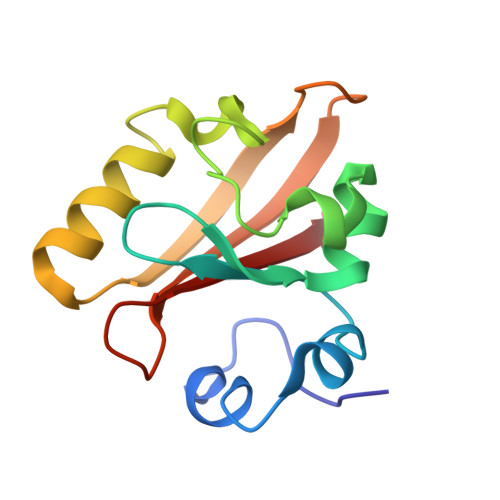
4I3A | pdb_00004i3a

Structures of PR1 and PR2 intermediates from time-resolved laue crystallography collected at 14ID-B, APS


Experimental Data Snapshot

  • Method: X-RAY DIFFRACTION
  • Resolution: 1.60 Å
  • R-Value Free: 
    0.349 (Depositor), 0.321 (DCC) 
  • R-Value Work: 
    0.298 (Depositor), 0.288 (DCC) 
  • R-Value Observed: 
    0.301 (Depositor) 

wwPDB Validation   3D Report Full Report


Ligand Structure Quality Assessment 

Created with Raphaël 2.3.0Worse 01 BetterLigand structure goodness of fit to experimental dataBest fitted HC4Click on this verticalbar to view details

This is version 1.2 of the entry. See complete history


Literature

Volume-conserving trans-cis isomerization pathways in photoactive yellow protein visualized by picosecond X-ray crystallography

Jung, Y.O.Lee, J.H.Kim, J.Schmidt, M.Moffat, K.Srajer, V.Ihee, H.

(2013) Nat Chem 5: 212-220

  • DOI: https://doi.org/10.1038/nchem.1565
  • Primary Citation of Related Structures:  
    3VE3, 3VE4, 4HY8, 4I38, 4I39, 4I3A, 4I3I, 4I3J

  • PubMed Abstract: 

    Trans-to-cis isomerization, the key reaction in photoactive proteins, usually cannot occur through the standard one-bond-flip mechanism. Owing to spatial constraints imposed by a protein environment, isomerization probably proceeds through a volume-conserving mechanism in which highly choreographed atomic motions are expected, the details of which have not yet been observed directly. Here we employ time-resolved X-ray crystallography to visualize structurally the isomerization of the p-coumaric acid chromophore in photoactive yellow protein with a time resolution of 100 ps and a spatial resolution of 1.6 Å. The structure of the earliest intermediate (I(T)) resembles a highly strained transition state in which the torsion angle is located halfway between the trans- and cis-isomers. The reaction trajectory of I(T) bifurcates into two structurally distinct cis intermediates via hula-twist and bicycle-pedal pathways. The bifurcating reaction pathways can be controlled by weakening the hydrogen bond between the chromophore and an adjacent residue through E46Q mutation, which switches off the bicycle-pedal pathway.


  • Organizational Affiliation

    Center for Nanomaterials and Chemical Reactions, Institute for Basic Science, Daejeon 305-701, Republic of Korea.


Macromolecules
Find similar proteins by:  (by identity cutoff)  |  3D Structure
Entity ID: 1
MoleculeChains Sequence LengthOrganismDetailsImage
Photoactive yellow protein125Halorhodospira halophilaMutation(s): 0 
Gene Names: pyp
UniProt
Find proteins for P16113 (Halorhodospira halophila)
Explore P16113 
Go to UniProtKB:  P16113
Entity Groups  
Sequence Clusters30% Identity50% Identity70% Identity90% Identity95% Identity100% Identity
UniProt GroupP16113
Sequence Annotations
Expand
  • Reference Sequence
Small Molecules
Ligands 1 Unique
IDChains Name / Formula / InChI Key2D Diagram3D Interactions
HC4
Query on HC4

Download Ideal Coordinates CCD File 
B [auth A]4'-HYDROXYCINNAMIC ACID
C9 H8 O3
NGSWKAQJJWESNS-ZZXKWVIFSA-N
Experimental Data & Validation

Experimental Data

  • Method: X-RAY DIFFRACTION
  • Resolution: 1.60 Å
  • R-Value Free:  0.349 (Depositor), 0.321 (DCC) 
  • R-Value Work:  0.298 (Depositor), 0.288 (DCC) 
  • R-Value Observed: 0.301 (Depositor) 
Space Group: P 63
Unit Cell:
Length ( Å )Angle ( ˚ )
a = 66.833α = 90
b = 66.833β = 90
c = 40.947γ = 120
Software Package:
Software NamePurpose
SHELXrefinement
PDB_EXTRACTdata extraction
LaueViewdata scaling
LaueViewdata reduction
XFITdata reduction
SHELXL-97refinement

Structure Validation

View Full Validation Report


Ligand Structure Quality Assessment 

Created with Raphaël 2.3.0Worse 01 BetterLigand structure goodness of fit to experimental dataBest fitted HC4Click on this verticalbar to view details

Entry History 

Deposition Data

Revision History  (Full details and data files)

  • Version 1.0: 2013-03-20
    Type: Initial release
  • Version 1.1: 2017-11-15
    Changes: Refinement description
  • Version 1.2: 2025-03-26
    Changes: Data collection, Database references, Derived calculations, Structure summary