4HYQ

Crystal structure of phospholipase A1 from Streptomyces albidoflavus NA297


Experimental Data Snapshot

  • Method: X-RAY DIFFRACTION
  • Resolution: 1.75 Å
  • R-Value Free: 0.223 
  • R-Value Work: 0.186 
  • R-Value Observed: 0.186 

wwPDB Validation   3D Report Full Report


This is version 1.1 of the entry. See complete history


Literature

Crystal structure of phospholipase A1 from Streptomyces albidoflavus NA297

Murayama, K.Kano, K.Matsumoto, Y.Sugimori, D.

(2013) J Struct Biol 182: 192-196

  • DOI: https://doi.org/10.1016/j.jsb.2013.02.003
  • Primary Citation of Related Structures:  
    4HYQ

  • PubMed Abstract: 

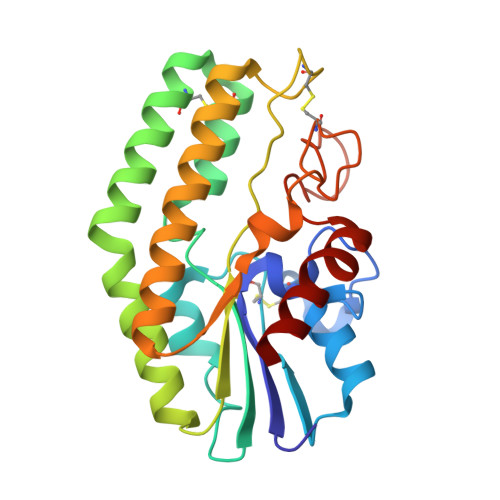
    The metal-independent lipase from Streptomyces albidoflavus NA297 (SaPLA1) is a phospholipase A1 as it preferentially hydrolyzes the sn-1 acyl ester in glycerophospholipids, yielding a fatty acid and 2-acyl-lysophospholipid. The molecular mechanism underlying the substrate binding by SaPLA1 is currently unknown. In this study, the crystal structure of SaPLA1 was determined at 1.75Å resolutions by molecular replacement. A structural similarity search indicated the highest structural similarity to an esterase from Streptomyces scabies, followed by GDSL family enzymes. The SaPLA1 active site is composed of a Ser-His dyad (Ser11 and His218), whereby stabilization of the imidazole is provided by the main-chain carbonyl oxygen of Ser216, a common variation of the catalytic triad in many serine hydrolases, where this carbonyl maintains the orientation of the active site histidine residue. The hydrophobic pocket and cleft for lipid binding are adjacent to the active site, and are approximately 13-15Å deep and 14-16Å long. A partial polyethylene glycol structure was found in this hydrophobic pocket.


  • Organizational Affiliation

    Graduate School of Biomedical Engineering, Tohoku University, Seiryo 2-1, Aoba, Sendai 980-8575, Japan. kmura@bme.tohoku.ac.jp


Macromolecules
Find similar proteins by:  (by identity cutoff)  |  3D Structure
Entity ID: 1
MoleculeChains Sequence LengthOrganismDetailsImage
phospholipase A1236Streptomyces albidoflavusMutation(s): 0 
EC: 3.1.1.32
Membrane Entity: Yes 
UniProt
Find proteins for K0J3J2 (Streptomyces albidoflavus)
Explore K0J3J2 
Go to UniProtKB:  K0J3J2
Entity Groups  
Sequence Clusters30% Identity50% Identity70% Identity90% Identity95% Identity100% Identity
UniProt GroupK0J3J2
Sequence Annotations
Expand
  • Reference Sequence
Small Molecules
Ligands 1 Unique
IDChains Name / Formula / InChI Key2D Diagram3D Interactions
1PE
Query on 1PE

Download Ideal Coordinates CCD File 
B [auth A]PENTAETHYLENE GLYCOL
C10 H22 O6
JLFNLZLINWHATN-UHFFFAOYSA-N
Experimental Data & Validation

Experimental Data

  • Method: X-RAY DIFFRACTION
  • Resolution: 1.75 Å
  • R-Value Free: 0.223 
  • R-Value Work: 0.186 
  • R-Value Observed: 0.186 
  • Space Group: P 31 2 1
Unit Cell:
Length ( Å )Angle ( ˚ )
a = 67.55α = 90
b = 67.55β = 90
c = 96.112γ = 120
Software Package:
Software NamePurpose
HKL-2000data collection
MOLREPphasing
CNSrefinement
HKL-2000data reduction
HKL-2000data scaling

Structure Validation

View Full Validation Report


Entry History 

Deposition Data

Revision History  (Full details and data files)

  • Version 1.0: 2013-05-29
    Type: Initial release
  • Version 1.1: 2023-11-08
    Changes: Data collection, Database references, Derived calculations, Refinement description