4HX9 | pdb_00004hx9

Designed Phosphodeoxyribosyltransferase


Experimental Data Snapshot

  • Method: X-RAY DIFFRACTION
  • Resolution: 2.68 Å
  • R-Value Free: 
    0.250 (Depositor), 0.242 (DCC) 
  • R-Value Work: 
    0.195 (Depositor), 0.192 (DCC) 
  • R-Value Observed: 
    0.198 (Depositor) 

Starting Model: experimental
View more details

wwPDB Validation 3D Report Full Report

Validation slider image for 4HX9

This is version 1.3 of the entry. See complete history

Literature

Phosphodeoxyribosyltransferases, designed enzymes for deoxyribonucleotides synthesis.

Kaminski, P.A.Labesse, G.

(2013) J Biological Chem 288: 6534-6541

  • DOI: https://doi.org/10.1074/jbc.M112.446492
  • Primary Citation Related Structures: 
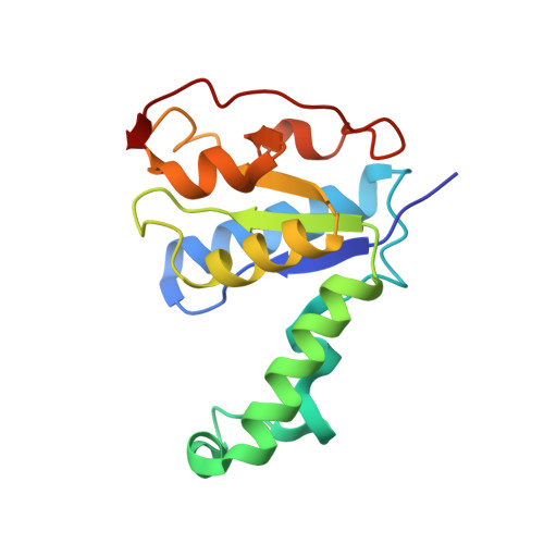
    4HX9

  • PubMed Abstract: 

    A large number of nucleoside analogues and 2'-deoxynucleoside triphosphates (dNTP) have been synthesized to interfere with DNA metabolism. However, in vivo the concentration and phosphorylation of these analogues are key limiting factors. In this context, we designed enzymes to switch nucleobases attached to a deoxyribose monophosphate. Active chimeras were made from two distantly related enzymes: a nucleoside deoxyribosyltransferase from lactobacilli and a 5'-monophosphate-2'-deoxyribonucleoside hydrolase from rat. Then their unprecedented activity was further extended to deoxyribose triphosphate, and in vitro biosyntheses could be successfully performed with several base analogues. These new enzymes provide new tools to synthesize dNTP analogues and to deliver them into cells.


  • Organizational Affiliation
    • Institut Pasteur, Unité de Chimie et Biocatalyse, CNRS, UMR 3523, 75724 Paris cedex 15, France. pierre-alexandre.kaminski@pasteur.fr

Macromolecule Content 

  • Total Structure Weight: 146.7 kDa 
  • Atom Count: 10,448 
  • Modeled Residue Count: 1,245 
  • Deposited Residue Count: 1,256 
  • Unique protein chains: 1

Macromolecules

Find similar proteins by:|  3D Structure
Entity ID: 1
MoleculeChains  Sequence LengthOrganismDetailsImage
Nucleoside deoxyribosyltransferase157Lactobacillus leichmanniiMutation(s): 5 
Gene Names: ntd
EC: 2.4.2.6
UniProt
Find proteins for Q9R5V5 (Lactobacillus leichmannii)
Explore Q9R5V5 
Go to UniProtKB:  Q9R5V5
Entity Groups
Sequence Clusters30% Identity50% Identity70% Identity90% Identity95% Identity100% Identity
UniProt GroupQ9R5V5
Sequence Annotations
Expand
Reference Sequence

Small Molecules

Ligands 2 Unique
IDChains Name / Formula / InChI Key2D Diagram3D Interactions
PG4

Query on PG4


Download:Ideal Coordinates CCD File
K [auth A]
M [auth B]
R [auth F]
S [auth F]
W [auth D]
K [auth A],
M [auth B],
R [auth F],
S [auth F],
W [auth D],
X [auth D]
TETRAETHYLENE GLYCOL
C8 H18 O5
UWHCKJMYHZGTIT-UHFFFAOYSA-N
SO4

Query on SO4


Download:Ideal Coordinates CCD File
AA [auth H]
BA [auth H]
I [auth A]
J [auth A]
L [auth B]
AA [auth H],
BA [auth H],
I [auth A],
J [auth A],
L [auth B],
N [auth E],
O [auth E],
P [auth F],
Q [auth F],
T [auth C],
U [auth D],
V [auth D],
Y [auth G],
Z [auth G]
SULFATE ION
O4 S
QAOWNCQODCNURD-UHFFFAOYSA-L

Experimental Data & Validation

Experimental Data

  • Method: X-RAY DIFFRACTION
  • Resolution: 2.68 Å
  • R-Value Free:  0.250 (Depositor), 0.242 (DCC) 
  • R-Value Work:  0.195 (Depositor), 0.192 (DCC) 
  • R-Value Observed: 0.198 (Depositor) 
Space Group: I 21 3
Unit Cell:
Length ( Å )Angle ( ˚ )
a = 218.454α = 90
b = 218.454β = 90
c = 218.454γ = 90
Software Package:
Software NamePurpose
PROTEUM PLUSdata collection
AMoREphasing
REFMACrefinement
MOSFLMdata reduction
SCALAdata scaling

Structure Validation

View Full Validation Report


Entry History 

Deposition Data

Revision History  (Full details and data files)

  • Version 1.0: 2013-01-16
    Type: Initial release
  • Version 1.1: 2013-02-13
    Changes: Database references
  • Version 1.2: 2013-03-20
    Changes: Database references
  • Version 1.3: 2023-09-20
    Changes: Data collection, Database references, Derived calculations, Refinement description