4HW9 | pdb_00004hw9

Crystal Structure of Helicobacter pylori MscS (Closed State)


Experimental Data Snapshot

  • Method: X-RAY DIFFRACTION
  • Resolution: 4.14 Å
  • R-Value Free: 
    0.304 (Depositor), 0.307 (DCC) 
  • R-Value Work: 
    0.244 (Depositor), 0.255 (DCC) 
  • R-Value Observed: 
    0.247 (Depositor) 

wwPDB Validation 3D Report Full Report

Validation slider image for 4HW9

This is version 1.3 of the entry. See complete history

Literature

Open and shut: crystal structures of the dodecylmaltoside solubilized mechanosensitive channel of small conductance from Escherichia coli and Helicobacter pylori at 4.4 angstrom and 4.1 angstrom resolutions.

Lai, J.Y.Poon, Y.S.Kaiser, J.T.Rees, D.C.

(2013) Protein Sci 22: 502-509

  • DOI: https://doi.org/10.1002/pro.2222
  • Primary Citation Related Structures: 
    4HW9, 4HWA

  • PubMed Abstract: 

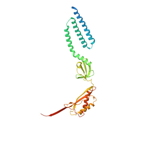
    The mechanosensitive channel of small conductance (MscS) contributes to the survival of bacteria during osmotic downshock by transiently opening large diameter pores for the efflux of cellular contents before the membrane ruptures. Two crystal structures of the Escherichia coli MscS are currently available, the wild type protein in a nonconducting state at 3.7 Å resolution (Bass et al., Science 2002; 298:1582-1587) and the Ala106Val variant in an open state at 3.45 Å resolution (Wang et al., Science 2008; 321:1179-1183). Both structures used protein solubilized in the detergent fos-choline-14. We report here crystal structures of MscS from E. coli and Helicobacter pylori solubilized in the detergent β-dodecylmaltoside at resolutions of 4.4 and 4.2 Å, respectively. While the cytoplasmic domains are unchanged in these structures, distinct conformations of the transmembrane domains are observed. Intriguingly, β-dodecylmaltoside solubilized wild type E. coli MscS adopts the open state structure of A106V E. coli MscS, while H. pylori MscS resembles the nonconducting state structure observed for fos-choline-14 solubilized E. coli MscS. These results highlight the sensitivity of membrane protein conformational equilibria to variations in detergent, crystallization conditions, and protein sequence.


  • Organizational Affiliation
    • Division of Chemistry and Chemical Engineering, Howard Hughes Medical Institute, California Institute of Technology, Pasadena, California 91125, USA.

Macromolecule Content 

  • Total Structure Weight: 240.77 kDa 
  • Atom Count: 13,797 
  • Modeled Residue Count: 1,771 
  • Deposited Residue Count: 2,163 
  • Unique protein chains: 1

Macromolecules

Find similar proteins by:|  3D Structure
Entity ID: 1
MoleculeChains  Sequence LengthOrganismDetailsImage
Mechanosensitive channel MscS
A, B, C, D, E
A, B, C, D, E, F, G
309Helicobacter pyloriMutation(s): 0 
Gene Names: MscS
Membrane Entity: Yes 
UniProt
Find proteins for E8QGV2 (Helicobacter pylori (strain India7))
Explore E8QGV2 
Go to UniProtKB:  E8QGV2
Entity Groups
Sequence Clusters30% Identity50% Identity70% Identity90% Identity95% Identity100% Identity
UniProt GroupE8QGV2
Sequence Annotations
Expand
Reference Sequence

Experimental Data & Validation

Experimental Data

  • Method: X-RAY DIFFRACTION
  • Resolution: 4.14 Å
  • R-Value Free:  0.304 (Depositor), 0.307 (DCC) 
  • R-Value Work:  0.244 (Depositor), 0.255 (DCC) 
  • R-Value Observed: 0.247 (Depositor) 
Space Group: P 21 21 21
Unit Cell:
Length ( Å )Angle ( ˚ )
a = 113.92α = 90
b = 143.1β = 90
c = 178.37γ = 90
Software Package:
Software NamePurpose
Blu-Icedata collection
PHASERphasing
PHENIXrefinement
XDSdata reduction
SCALAdata scaling

Structure Validation

View Full Validation Report


Entry History 

Deposition Data

Revision History  (Full details and data files)

  • Version 1.0: 2013-02-13
    Type: Initial release
  • Version 1.1: 2013-08-28
    Changes: Database references
  • Version 1.2: 2018-01-24
    Changes: Structure summary
  • Version 1.3: 2024-02-28
    Changes: Data collection, Database references, Refinement description