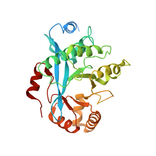
Structures of the catalytic EAL domain of the Escherichia coli direct oxygen sensor.
Tarnawski, M., Barends, T.R., Hartmann, E., Schlichting, I.(2013) Acta Crystallogr D Biol Crystallogr 69: 1045-1053
- PubMed: 23695249
- DOI: https://doi.org/10.1107/S0907444913004423
- Primary Citation of Related Structures:
4HU3, 4HU4 - PubMed Abstract:
The direct oxygen sensor DosP is a multidomain protein that contains a gas-sensing haem domain and an EAL effector domain. EAL domains are omnipresent signal transduction domains in bacteria. Many EAL domains are active phosphodiesterases and are involved in breakdown of the ubiquitous bacterial second messenger cyclic di-GMP. Despite a great deal of information on the functional and structural aspects of active and inactive EAL domains, little is known about the structural basis of their regulation by their associated sensory domains. Here, two crystal structures of the Escherichia coli DosP EAL domain derived from cubic and monoclinic crystal forms that were obtained under tartrate and PEG conditions, respectively, are described. Both of the structures display the typical TIM (triosephosphate isomerase) barrel fold with one antiparallel β-strand. However, unlike other EAL structures, access to the active site in DosP EAL is sterically restricted by the presence of a short helical stretch (Ser637-Ala-Leu-His640) in loop L3 between strand β3 and helix α3. This element, together with an unordered fragment, replaces the short α-helix (named α5 in Tbd1265 EAL) that is found in other EAL-domain structures. Since DosP EAL is an active c-di-GMP phosphodiesterase, the observed inactive conformation is suggested to be of functional relevance for the regulation mechanism of DosP.
- Department of Biomolecular Mechanisms, Max Planck Institute for Medical Research, Jahnstrasse 29, Heidelberg, Germany.
Organizational Affiliation:


