Role of polarity of the distal pocket in the control of inhibitor binding in dehaloperoxidase-hemoglobin.
Plummer, A., Thompson, M.K., Franzen, S.(2013) Biochemistry 52: 2218-2227
- PubMed: 23480794 Search on PubMed
- DOI: https://doi.org/10.1021/bi301509r
- Primary Citation Related Structures:
4HSW, 4HSX - PubMed Abstract:
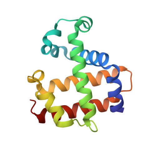
Dehaloperoxidase (DHP A), a unique multifunctional enzyme, from the marine annelid Amphitrite ornata dehalogenates 2,4,6-tribromophenol to form 2,6-dibromo-1,4-benzoquinone. The catalytic cycle of DHP is similar to that of horseradish peroxidase (HRP), involving a high-valent ferryl heme and two single-electron transfers from the aromatic substrate to the enzyme. Like HRP, DHP has been investigated as a potential bioremediation enzyme. However, DHP fails as a bioremediation enzyme because, unlike HRP, it has an internal binding cavity on the distal side of the heme capable of accommodating p-bromophenols, which act as an inhibitor of peroxidase function. Blocking internal binding in DHP may be the key to allowing the enzyme to function effectively as a peroxidase on the full range of halogenated phenols. The distal cavity of DHP is surrounded by several hydrophobic amino acids that stabilize internal binding of the monohalogenated phenols, including a leucine residue near the back edge of the heme (L100). We have expressed the L100F, L100Q, L100T, and L100V mutants of DHP in an effort to prevent internal binding and thereby convert the inhibitors into substrates. Kinetic assays and resonance Raman indicate that the peroxidase activity of the L100T and L100F mutants is increased compared to that of native DHP in the presence of 4-bromophenol (4-BP), suggesting a reduction in the inhibitor binding constant. In addition, the X-ray crystal structure of L100F clearly indicates a reduced occupancy of the 4-BP inhibitor in the distal cavity of DHP. However, at the same time, the L100F structure reveals that steric interference alone is insufficient to exclude the inhibitor. Instead, the comparison of L100T and isosteric L100V reveals that an increase in polarity plays a decisive role in excluding the inhibitor from the distal binding pocket.
- Department of Chemistry, North Carolina State University, Raleigh, NC 27695, USA.
Organizational Affiliation: