4HRT

Scapharca tetrameric hemoglobin, unliganded


Experimental Data Snapshot

  • Method: X-RAY DIFFRACTION
  • Resolution: 1.46 Å
  • R-Value Free: 0.175 
  • R-Value Work: 0.133 
  • R-Value Observed: 0.135 

wwPDB Validation   3D Report Full Report


Ligand Structure Quality Assessment 


This is version 1.3 of the entry. See complete history


Literature

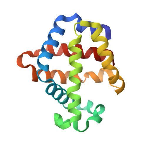
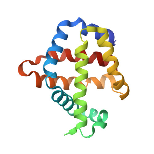
Tertiary and Quaternary Allostery in Tetrameric Hemoglobin from Scapharca inaequivalvis.

Ronda, L.Bettati, S.Henry, E.R.Kashav, T.Sanders, J.M.Royer, W.E.Mozzarelli, A.

(2013) Biochemistry 52: 2108-2117

  • DOI: https://doi.org/10.1021/bi301620x
  • Primary Citation of Related Structures:  
    4HRR, 4HRT

  • PubMed Abstract: 

    The clam Scapharca inaequivalvis possesses two cooperative oxygen binding hemoglobins in its red cells: a homodimeric HbI and a heterotetrameric A2B2 HbII. Each AB dimeric half of HbII is assembled in a manner very similar to that of the well-studied HbI. This study presents crystal structures of HbII along with oxygen binding data both in the crystalline state and in wet nanoporous silica gels. Despite very similar ligand-linked structural transitions observed in HbI and HbII crystals, HbII in the crystal or encapsulated in silica gels apparently exhibits minimal cooperativity in oxygen binding, in contrast with the full cooperativity exhibited by HbI crystals. However, oxygen binding curves in the crystal indicate the presence of a significant functional inequivalence of A and B chains. When this inequivalence is taken into account, both crystal and R state gel functional data are consistent with the conservation of a tertiary contribution to cooperative oxygen binding, quantitatively similar to that measured for HbI, and are in keeping with the structural information. Furthermore, our results indicate that to fully express cooperative ligand binding, HbII requires quaternary transitions hampered by crystal lattice and gel encapsulation, revealing greater complexity in cooperative function than the direct communication across a dimeric interface observed in HbI.


  • Organizational Affiliation

    Department of Pharmacy, University of Parma , Parco Area delle Scienze, 23/A, 43124 Parma, Italy.


Macromolecules
Find similar proteins by:  (by identity cutoff)  |  3D Structure
Entity ID: 1
MoleculeChains Sequence LengthOrganismDetailsImage
Globin-2 A chain
A, C, E, G
150Anadara inaequivalvisMutation(s): 1 
UniProt
Find proteins for P14821 (Anadara inaequivalvis)
Explore P14821 
Go to UniProtKB:  P14821
Entity Groups  
Sequence Clusters30% Identity50% Identity70% Identity90% Identity95% Identity100% Identity
UniProt GroupP14821
Sequence Annotations
Expand
  • Reference Sequence
Find similar proteins by:  (by identity cutoff)  |  3D Structure
Entity ID: 2
MoleculeChains Sequence LengthOrganismDetailsImage
Hemoglobin B chain
B, D, F, H
152Anadara inaequivalvisMutation(s): 1 
UniProt
Find proteins for P14822 (Anadara inaequivalvis)
Explore P14822 
Go to UniProtKB:  P14822
Entity Groups  
Sequence Clusters30% Identity50% Identity70% Identity90% Identity95% Identity100% Identity
UniProt GroupP14822
Sequence Annotations
Expand
  • Reference Sequence
Small Molecules
Ligands 2 Unique
IDChains Name / Formula / InChI Key2D Diagram3D Interactions
HEM
Query on HEM

Download Ideal Coordinates CCD File 
I [auth A]
J [auth B]
K [auth C]
L [auth D]
M [auth E]
I [auth A],
J [auth B],
K [auth C],
L [auth D],
M [auth E],
O [auth F],
P [auth G],
Q [auth H]
PROTOPORPHYRIN IX CONTAINING FE
C34 H32 Fe N4 O4
KABFMIBPWCXCRK-RGGAHWMASA-L
PO4
Query on PO4

Download Ideal Coordinates CCD File 
N [auth E]PHOSPHATE ION
O4 P
NBIIXXVUZAFLBC-UHFFFAOYSA-K
Experimental Data & Validation

Experimental Data

  • Method: X-RAY DIFFRACTION
  • Resolution: 1.46 Å
  • R-Value Free: 0.175 
  • R-Value Work: 0.133 
  • R-Value Observed: 0.135 
  • Space Group: P 2 2 21
Unit Cell:
Length ( Å )Angle ( ˚ )
a = 94.353α = 90
b = 100.958β = 90
c = 127.165γ = 90
Software Package:
Software NamePurpose
ADSCdata collection
AMoREphasing
REFMACrefinement
HKL-2000data reduction
HKL-2000data scaling

Structure Validation

View Full Validation Report


Ligand Structure Quality Assessment 


Entry History 

Deposition Data

  • Released Date: 2012-12-12 
  • Deposition Author(s): Royer, W.E.

Revision History  (Full details and data files)

  • Version 1.0: 2012-12-12
    Type: Initial release
  • Version 1.1: 2013-04-10
    Changes: Database references
  • Version 1.2: 2014-01-08
    Changes: Database references
  • Version 1.3: 2023-09-20
    Changes: Data collection, Database references, Derived calculations, Refinement description