


The chloride binding site of Neurotransmitter Sodium Symporters
Kantcheva, A.K., Quick, M., Shi, L., Winther, A.M.L., Stolzenberg, S., Weinstein, H., Javitch, J.A., Nissen, P.(2013) Proc Natl Acad Sci U S A
Experimental Data Snapshot
(2013) Proc Natl Acad Sci U S A
Entity ID: 1 | |||||
|---|---|---|---|---|---|
| Molecule | Chains | Sequence Length | Organism | Details | Image |
| Transporter | 515 | Aquifex aeolicus VF5 | Mutation(s): 1 Gene Names: snf, aq_2077 Membrane Entity: Yes | 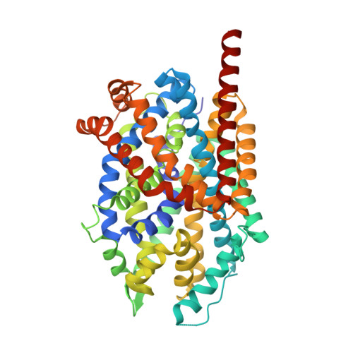 | |
UniProt | |||||
Entity Groups | |||||
| Sequence Clusters | 30% Identity50% Identity70% Identity90% Identity95% Identity100% Identity | ||||
| UniProt Group | O67854 | ||||
Sequence AnnotationsExpand | |||||
Reference Sequence | |||||
| Ligands 4 Unique | |||||
|---|---|---|---|---|---|
| ID | Chains | Name / Formula / InChI Key | 2D Diagram | 3D Interactions | |
| BOG Download:Ideal Coordinates CCD File | F [auth A], G [auth A], H [auth A], I [auth A], J [auth A] | octyl beta-D-glucopyranoside C14 H28 O6 HEGSGKPQLMEBJL-RKQHYHRCSA-N |  | ||
| LEU Download:Ideal Coordinates CCD File | B [auth A] | LEUCINE C6 H13 N O2 ROHFNLRQFUQHCH-YFKPBYRVSA-N |  | ||
| CL Download:Ideal Coordinates CCD File | E [auth A] | CHLORIDE ION Cl VEXZGXHMUGYJMC-UHFFFAOYSA-M |  | ||
| NA Download:Ideal Coordinates CCD File | C [auth A], D [auth A] | SODIUM ION Na FKNQFGJONOIPTF-UHFFFAOYSA-N |  | ||
| Length ( Å ) | Angle ( ˚ ) |
|---|---|
| a = 87.58 | α = 90 |
| b = 87.48 | β = 95 |
| c = 80.86 | γ = 90 |
| Software Name | Purpose |
|---|---|
| MAR345 | data collection |
| PHASER | phasing |
| PHENIX | refinement |
| XDS | data reduction |
| XSCALE | data scaling |


