


Crystal structure of Serine acetyltransferase from Vibrio cholerae O1 biovar El Tor N16961.
F Tarique, K., Abdul Rehman, S.A., Gourinath, S.To be published.
Experimental Data Snapshot
Starting Model: experimental
View more details
Entity ID: 1 | |||||
|---|---|---|---|---|---|
| Molecule | Chains | Sequence Length | Organism | Details | Image |
| Serine acetyltransferase | 281 | Vibrio cholerae O1 biovar El Tor str. N16961 | Mutation(s): 0 Gene Names: serine acetyltransferase (EC:2.3.1.30), VC_2649 EC: 2.3.1.30 | 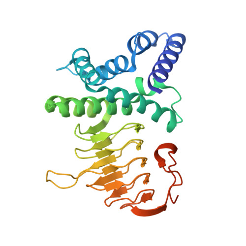 | |
UniProt | |||||
Entity Groups | |||||
| Sequence Clusters | 30% Identity50% Identity70% Identity90% Identity95% Identity100% Identity | ||||
| UniProt Group | Q9KNT2 | ||||
Sequence AnnotationsExpand | |||||
Reference Sequence | |||||
| Ligands 3 Unique | |||||
|---|---|---|---|---|---|
| ID | Chains | Name / Formula / InChI Key | 2D Diagram | 3D Interactions | |
| ARG Download:Ideal Coordinates CCD File | E [auth A], G [auth B], I [auth C] | ARGININE C6 H15 N4 O2 ODKSFYDXXFIFQN-BYPYZUCNSA-O |  | ||
| CYS Download:Ideal Coordinates CCD File | D [auth A], F [auth A], H [auth B] | CYSTEINE C3 H7 N O2 S XUJNEKJLAYXESH-REOHCLBHSA-N |  | ||
| NA Download:Ideal Coordinates CCD File | J [auth C] | SODIUM ION Na FKNQFGJONOIPTF-UHFFFAOYSA-N |  | ||
| Length ( Å ) | Angle ( ˚ ) |
|---|---|
| a = 144.04 | α = 90 |
| b = 75.04 | β = 89.04 |
| c = 73.43 | γ = 90 |
| Software Name | Purpose |
|---|---|
| DNA | data collection |
| PHASER | phasing |
| REFMAC | refinement |
| HKL-2000 | data reduction |
| HKL-2000 | data scaling |


