


Crystal structure of wild type protective antigen to 1.62 A (pH 7.5)
Sorrell, F.J., Rodgers, H.F., Baker, P.J., Chen, B.To be published.
Experimental Data Snapshot
Starting Model: experimental
View more details
wwPDB Validation 3D Report Full Report

Entity ID: 1 | |||||
|---|---|---|---|---|---|
| Molecule | Chains | Sequence Length | Organism | Details | Image |
| Protective antigen | 735 | Bacillus anthracis | Mutation(s): 0 Gene Names: pagA, pag, pXO1-110, BXA0164, GBAA_pXO1_0164 | 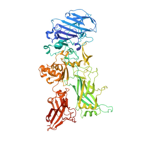 | |
UniProt | |||||
Entity Groups | |||||
| Sequence Clusters | 30% Identity50% Identity70% Identity90% Identity95% Identity100% Identity | ||||
| UniProt Group | P13423 | ||||
Sequence AnnotationsExpand | |||||
Reference Sequence | |||||
| Ligands 2 Unique | |||||
|---|---|---|---|---|---|
| ID | Chains | Name / Formula / InChI Key | 2D Diagram | 3D Interactions | |
| EDO Download:Ideal Coordinates CCD File | B [auth A], C [auth A], D [auth A], E [auth A] | 1,2-ETHANEDIOL C2 H6 O2 LYCAIKOWRPUZTN-UHFFFAOYSA-N |  | ||
| CA Download:Ideal Coordinates CCD File | F [auth A], G [auth A] | CALCIUM ION Ca BHPQYMZQTOCNFJ-UHFFFAOYSA-N |  | ||
| Length ( Å ) | Angle ( ˚ ) |
|---|---|
| a = 74.05 | α = 90 |
| b = 94.09 | β = 90 |
| c = 117.39 | γ = 90 |
| Software Name | Purpose |
|---|---|
| REFMAC | refinement |
| PDB_EXTRACT | data extraction |
| ADSC | data collection |
| XDS | data reduction |
| XSCALE | data scaling |
| PHASER | phasing |


