4H1E | pdb_00004h1e

Structure of BACE-1 Bound to (7aR)-6-benzoyl-7a-(4-(3-cyanophenyl)thiophen-2-yl)-3-methyl-4-oxohexahydro-1H-pyrrolo[3,4-d]pyrimidin-2(3H)-iminium


Experimental Data Snapshot

  • Method: X-RAY DIFFRACTION
  • Resolution: 1.90 Å
  • R-Value Free: 
    0.297 (Depositor), 0.286 (DCC) 
  • R-Value Work: 
    0.260 (Depositor), 0.251 (DCC) 
  • R-Value Observed: 
    0.262 (Depositor) 

Starting Model: experimental
View more details

wwPDB Validation 3D Report Full Report

Validation slider image for 4H1E

Ligand Structure Quality Assessment 


This is version 1.3 of the entry. See complete history

Literature

Design and Validation of Bicyclic Iminopyrimidinones As Beta Amyloid Cleaving Enzyme-1 (BACE1) Inhibitors: Conformational Constraint to Favor a Bioactive Conformation.

Mandal, M.Zhu, Z.Cumming, J.N.Liu, X.Strickland, C.Mazzola, R.D.Caldwell, J.P.Leach, P.Grzelak, M.Hyde, L.Zhang, Q.Terracina, G.Zhang, L.Chen, X.Kuvelkar, R.Kennedy, M.E.Favreau, L.Cox, K.Orth, P.Buevich, A.Voigt, J.Wang, H.Kazakevich, I.McKittrick, B.A.Greenlee, W.Parker, E.M.Stamford, A.W.

(2012) J Med Chem 55: 9331-9345

  • DOI: https://doi.org/10.1021/jm301039c
  • Primary Citation Related Structures: 
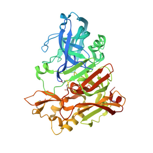
    4H1E, 4H3F, 4H3G, 4H3I, 4H3J, 4HA5

  • PubMed Abstract: 

    On the basis of our observation that the biaryl substituent of iminopyrimidinone 7 must be in a pseudoaxial conformation to occupy the contiguous S1-S3 subsites of BACE1, we have designed a novel fused bicyclic iminopyrimidinone scaffold intended to favor this bioactive conformation. Strategic incorporation of a nitrogen atom in the new constrained ring allowed us to develop SAR around the S2' binding pocket and ultimately resulted in analogues with low nanomolar potency for BACE1. In particular, optimization of the prime side substituent led to major improvements in potency by displacement of two conserved water molecules from a region near S2'. Further optimization of the pharmacokinetic properties of this fused pyrrolidine series, in conjunction with facile access to a rat pharmacodynamic model, led to identification of compound 43, which is an orally active, brain penetrant inhibitor that reduces Aβ(40) in the plasma, CSF, and cortex of rats in a dose-dependent manner.


  • Organizational Affiliation
    • Department of Medicinal Chemistry, Merck Research Laboratories, 2015 Galloping Hill Road, Kenilworth, New Jersey 07033, United States. mihirbaran.mandal@merck.com

Macromolecule Content 

  • Total Structure Weight: 93.41 kDa 
  • Atom Count: 6,608 
  • Modeled Residue Count: 773 
  • Deposited Residue Count: 828 
  • Unique protein chains: 1

Macromolecules

Find similar proteins by:|  3D Structure
Entity ID: 1
MoleculeChains  Sequence LengthOrganismDetailsImage
Beta-secretase 1
A, B
414Homo sapiensMutation(s): 0 
Gene Names: BACEBACE1KIAA1149
EC: 3.4.23.46
UniProt & NIH Common Fund Data Resources
Find proteins for P56817 (Homo sapiens)
Explore P56817 
Go to UniProtKB:  P56817
PHAROS:  P56817
GTEx:  ENSG00000186318 
Entity Groups
Sequence Clusters30% Identity50% Identity70% Identity90% Identity95% Identity100% Identity
UniProt GroupP56817
Sequence Annotations
Expand
Reference Sequence

Small Molecules

Ligands 1 Unique
IDChains Name / Formula / InChI Key2D Diagram3D Interactions
10J

Query on 10J


Download:Ideal Coordinates CCD File
C [auth A],
D [auth B]
3-{5-[(2E,4aR,7aR)-6-benzoyl-2-imino-3-methyl-4-oxooctahydro-7aH-pyrrolo[3,4-d]pyrimidin-7a-yl]thiophen-3-yl}benzonitrile
C25 H21 N5 O2 S
POOXGNGZUXTIQE-CPJSRVTESA-N

Experimental Data & Validation

Experimental Data

  • Method: X-RAY DIFFRACTION
  • Resolution: 1.90 Å
  • R-Value Free:  0.297 (Depositor), 0.286 (DCC) 
  • R-Value Work:  0.260 (Depositor), 0.251 (DCC) 
  • R-Value Observed: 0.262 (Depositor) 
Space Group: P 21 21 21
Unit Cell:
Length ( Å )Angle ( ˚ )
a = 86.212α = 90
b = 89.936β = 90
c = 131.323γ = 90
Software Package:
Software NamePurpose
BUSTER-TNTrefinement
DENZOdata reduction
SCALEPACKdata scaling
PDB_EXTRACTdata extraction
CrystalCleardata collection
CNSphasing
BUSTERrefinement

Structure Validation

View Full Validation Report


Ligand Structure Quality Assessment 


Entry History 

Deposition Data

  • Released Date: 2012-10-17 
  • Deposition Author(s): Orth, P.

Revision History  (Full details and data files)

  • Version 1.0: 2012-10-17
    Type: Initial release
  • Version 1.1: 2012-11-21
    Changes: Database references
  • Version 1.2: 2023-09-13
    Changes: Data collection, Database references, Derived calculations, Refinement description
  • Version 1.3: 2024-11-20
    Changes: Structure summary