4GT7

An engineered disulfide bond reversibly traps the IgE-Fc3-4 in a closed, non-receptor binding conformation


Experimental Data Snapshot

  • Method: X-RAY DIFFRACTION
  • Resolution: 2.61 Å
  • R-Value Free: 0.253 
  • R-Value Work: 0.190 
  • R-Value Observed: 0.192 

wwPDB Validation   3D Report Full Report


This is version 2.0 of the entry. See complete history


Literature

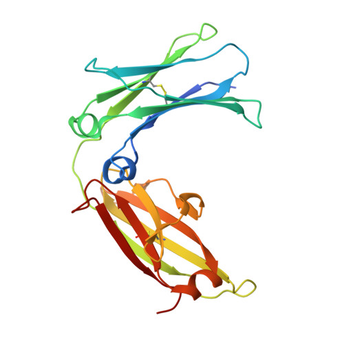
An engineered disulfide bond reversibly traps the IgE-Fc3-4 in a closed, nonreceptor binding conformation.

Wurzburg, B.A.Kim, B.Tarchevskaya, S.S.Eggel, A.Vogel, M.Jardetzky, T.S.

(2012) J Biol Chem 287: 36251-36257

  • DOI: https://doi.org/10.1074/jbc.M112.407502
  • Primary Citation of Related Structures:  
    4GT7

  • PubMed Abstract: 

    IgE antibodies interact with the high affinity IgE Fc receptor, FcεRI, and activate inflammatory pathways associated with the allergic response. The IgE-Fc region, comprising the C-terminal domains of the IgE heavy chain, binds FcεRI and can adopt different conformations ranging from a closed form incompatible with receptor binding to an open, receptor-bound state. A number of intermediate states are also observed in different IgE-Fc crystal forms. To further explore this apparent IgE-Fc conformational flexibility and to potentially trap a closed, inactive state, we generated a series of disulfide bond mutants. Here we describe the structure and biochemical properties of an IgE-Fc mutant that is trapped in the closed, non-receptor binding state via an engineered disulfide at residue 335 (Cys-335). Reduction of the disulfide at Cys-335 restores the ability of IgE-Fc to bind to its high affinity receptor, FcεRIα. The structure of the Cys-335 mutant shows that its conformation is within the range of previously observed, closed form IgE-Fc structures and that it retains the hydrophobic pocket found in the hinge region of the closed conformation. Locking the IgE-Fc into the closed state with the Cys-335 mutation does not affect binding of two other IgE-Fc ligands, omalizumab and DARPin E2_79, demonstrating selective blocking of the high affinity receptor binding.


  • Organizational Affiliation

    Department of Structural Biology, Stanford University School of Medicine, Stanford, California 94305, USA.


Macromolecules
Find similar proteins by:  (by identity cutoff)  |  3D Structure
Entity ID: 1
MoleculeChains Sequence LengthOrganismDetailsImage
Ig epsilon chain C region
A, B, C, D
221Homo sapiensMutation(s): 1 
Gene Names: IGHE
UniProt & NIH Common Fund Data Resources
Find proteins for P01854 (Homo sapiens)
Explore P01854 
Go to UniProtKB:  P01854
PHAROS:  P01854
GTEx:  ENSG00000211891 
Entity Groups  
Sequence Clusters30% Identity50% Identity70% Identity90% Identity95% Identity100% Identity
UniProt GroupP01854
Sequence Annotations
Expand
  • Reference Sequence
Oligosaccharides

Help

Entity ID: 2
MoleculeChains Length2D Diagram Glycosylation3D Interactions
alpha-D-mannopyranose-(1-3)-[alpha-D-mannopyranose-(1-6)]beta-D-mannopyranose-(1-4)-2-acetamido-2-deoxy-beta-D-glucopyranose-(1-4)-2-acetamido-2-deoxy-beta-D-glucopyranose
E, F, G
5N-Glycosylation
Glycosylation Resources
GlyTouCan:  G22768VO
GlyCosmos:  G22768VO
GlyGen:  G22768VO
Entity ID: 3
MoleculeChains Length2D Diagram Glycosylation3D Interactions
alpha-D-mannopyranose-(1-6)-beta-D-mannopyranose-(1-4)-2-acetamido-2-deoxy-beta-D-glucopyranose-(1-4)-2-acetamido-2-deoxy-beta-D-glucopyranose
H
4N-Glycosylation
Glycosylation Resources
GlyTouCan:  G22573RC
GlyCosmos:  G22573RC
GlyGen:  G22573RC
Experimental Data & Validation

Experimental Data

  • Method: X-RAY DIFFRACTION
  • Resolution: 2.61 Å
  • R-Value Free: 0.253 
  • R-Value Work: 0.190 
  • R-Value Observed: 0.192 
  • Space Group: P 1 21 1
Unit Cell:
Length ( Å )Angle ( ˚ )
a = 106.753α = 90
b = 104.807β = 94.51
c = 45.899γ = 90
Software Package:
Software NamePurpose
MAR345dtbdata collection
PHASERphasing
PHENIXrefinement
XDSdata reduction
XSCALEdata scaling

Structure Validation

View Full Validation Report


Entry History 

Deposition Data

Revision History  (Full details and data files)

  • Version 1.0: 2012-09-12
    Type: Initial release
  • Version 1.1: 2013-01-02
    Changes: Database references
  • Version 2.0: 2020-07-29
    Type: Remediation
    Reason: Carbohydrate remediation
    Changes: Atomic model, Data collection, Database references, Derived calculations, Structure summary