4GSP | pdb_00004gsp

RIBONUCLEASE T1 COMPLEXED WITH 2',3'-CGPS + 3'-GMP, 7 DAYS


Experimental Data Snapshot

  • Method: X-RAY DIFFRACTION
  • Resolution: 1.65 Å
  • R-Value Free: 
    0.197 (Depositor), 0.212 (DCC) 
  • R-Value Work: 
    0.159 (Depositor), 0.173 (DCC) 
  • R-Value Observed: 
    0.159 (Depositor) 

Starting Model: experimental
View more details

wwPDB Validation 3D Report Full Report

Validation slider image for 4GSP

Ligand Structure Quality Assessment 


This is version 1.4 of the entry. See complete history

Literature

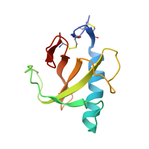
Hydrolysis of a slow cyclic thiophosphate substrate of RNase T1 analyzed by time-resolved crystallography.

Zegers, I.Loris, R.Dehollander, G.Fattah Haikal, A.Poortmans, F.Steyaert, J.Wyns, L.

(1998) Nat Struct Biol 5: 280-283

  • DOI: https://doi.org/10.1038/nsb0498-280
  • Primary Citation Related Structures: 
    1GSP, 2GSP, 3GSP, 4GSP, 5GSP, 6GSP, 7GSP

  • PubMed Abstract: 

    Here we present a time-resolved crystallographic analysis of the hydrolysis of exo (Sp) guanosine 2',3'-cyclophosphorothioate by RNase T1. The use of a slow substrate and fast crystallization methods made it possible to perform the study with conventional data-collection techniques. The results support the idea that the hydrolysis reaction proceeds through a mechanism that is the inverse of the transesterification reaction. In addition, the structures provide an explanation for the differential behavior of RNase T1 towards exo- and endo-cyclic thiophosphates.


  • Organizational Affiliation
    • Laboratorium voor Ultrastructuur, Vlaams Interuniversitair Instituut voor Biotechnologie, Vrije Universiteit Brussel, Belgium. igzegers@vub.ac.be

Macromolecule Content 

  • Total Structure Weight: 11.86 kDa 
  • Atom Count: 981 
  • Modeled Residue Count: 104 
  • Deposited Residue Count: 104 
  • Unique protein chains: 1

Macromolecules

Find similar proteins by:|  3D Structure
Entity ID: 1
MoleculeChains  Sequence LengthOrganismDetailsImage
RIBONUCLEASE T1104Aspergillus oryzaeMutation(s): 0 
EC: 3.1.27.3 (PDB Primary Data), 4.6.1.24 (UniProt)
UniProt
Find proteins for P00651 (Aspergillus oryzae (strain ATCC 42149 / RIB 40))
Explore P00651 
Go to UniProtKB:  P00651
Entity Groups
Sequence Clusters30% Identity50% Identity70% Identity90% Identity95% Identity100% Identity
UniProt GroupP00651
Sequence Annotations
Expand
Reference Sequence

Experimental Data & Validation

Experimental Data

  • Method: X-RAY DIFFRACTION
  • Resolution: 1.65 Å
  • R-Value Free:  0.197 (Depositor), 0.212 (DCC) 
  • R-Value Work:  0.159 (Depositor), 0.173 (DCC) 
  • R-Value Observed: 0.159 (Depositor) 
Space Group: P 21 21 21
Unit Cell:
Length ( Å )Angle ( ˚ )
a = 48.24α = 90
b = 51.1β = 90
c = 40.45γ = 90
Software Package:
Software NamePurpose
DENZOdata reduction
SCALEPACKdata scaling
X-PLORmodel building
X-PLORrefinement
X-PLORphasing

Structure Validation

View Full Validation Report


Ligand Structure Quality Assessment 


Entry History 

Deposition Data

Revision History  (Full details and data files)

  • Version 1.0: 1998-08-12
    Type: Initial release
  • Version 1.1: 2008-03-25
    Changes: Version format compliance
  • Version 1.2: 2011-07-13
    Changes: Version format compliance
  • Version 1.3: 2023-08-09
    Changes: Database references, Derived calculations, Refinement description
  • Version 1.4: 2024-10-30
    Changes: Data collection, Structure summary