4GM0

Crystal Structure of Benzoylformate Decarboxylase Mutant L403N


Experimental Data Snapshot

  • Method: X-RAY DIFFRACTION
  • Resolution: 1.07 Å
  • R-Value Free: 0.151 
  • R-Value Work: 0.137 
  • R-Value Observed: 0.138 

wwPDB Validation   3D Report Full Report


Ligand Structure Quality Assessment 


This is version 1.2 of the entry. See complete history


Literature

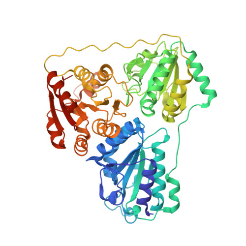
A bulky hydrophobic residue is not required to maintain the v-conformation of enzyme-bound thiamin diphosphate.

Andrews, F.H.Tom, A.R.Gunderman, P.R.Novak, W.R.McLeish, M.J.

(2013) Biochemistry 52: 3028-3030

  • DOI: https://doi.org/10.1021/bi400368j
  • Primary Citation of Related Structures:  
    4GG1, 4GM0, 4GM1, 4GM4, 4GP9, 4GPE, 4JD5

  • PubMed Abstract: 

    It is widely accepted that, in thiamin diphosphate (ThDP)-dependent enzymes, much of the rate acceleration is provided by the cofactor. Inter alia, the reactive conformation of ThDP, known as the V-conformation, has been attributed to the presence of a bulky hydrophobic residue located directly below the cofactor. Here we report the use of site-saturation mutagenesis to generate variants of this residue (Leu403) in benzoylformate decarboxylase. The observed 3 orders of magnitude range in k(cat)/K(m) values suggested that conformational changes in the cofactor could be influencing catalysis. However, X-ray structures of several variants were determined, and there was remarkably little change in ThDP conformation. Rather, it seemed that, once the V-conformation was attained, residue size and hydrophobicity were more important for enzyme activity.


  • Organizational Affiliation

    Department of Chemistry and Chemical Biology, Indiana University-Purdue University Indianapolis, Indianapolis, IN 46202, USA.


Macromolecules
Find similar proteins by:  (by identity cutoff)  |  3D Structure
Entity ID: 1
MoleculeChains Sequence LengthOrganismDetailsImage
Benzoylformate decarboxylase536Pseudomonas putidaMutation(s): 1 
Gene Names: mdlC
EC: 4.1.1.7
UniProt
Find proteins for P20906 (Pseudomonas putida)
Explore P20906 
Go to UniProtKB:  P20906
Entity Groups  
Sequence Clusters30% Identity50% Identity70% Identity90% Identity95% Identity100% Identity
UniProt GroupP20906
Sequence Annotations
Expand
  • Reference Sequence
Small Molecules
Ligands 4 Unique
IDChains Name / Formula / InChI Key2D Diagram3D Interactions
TZD
Query on TZD

Download Ideal Coordinates CCD File 
B [auth A]2-{3-[(4-AMINO-2-METHYLPYRIMIDIN-5-YL)METHYL]-4-METHYL-2-OXO-2,3-DIHYDRO-1,3-THIAZOL-5-YL}ETHYL TRIHYDROGEN DIPHOSPHATE
C12 H18 N4 O8 P2 S
ZGJUYGIRPQSCFA-UHFFFAOYSA-N
GOL
Query on GOL

Download Ideal Coordinates CCD File 
D [auth A],
E [auth A]
GLYCEROL
C3 H8 O3
PEDCQBHIVMGVHV-UHFFFAOYSA-N
CA
Query on CA

Download Ideal Coordinates CCD File 
C [auth A],
G [auth A]
CALCIUM ION
Ca
BHPQYMZQTOCNFJ-UHFFFAOYSA-N
NA
Query on NA

Download Ideal Coordinates CCD File 
F [auth A]SODIUM ION
Na
FKNQFGJONOIPTF-UHFFFAOYSA-N
Experimental Data & Validation

Experimental Data

  • Method: X-RAY DIFFRACTION
  • Resolution: 1.07 Å
  • R-Value Free: 0.151 
  • R-Value Work: 0.137 
  • R-Value Observed: 0.138 
  • Space Group: I 2 2 2
Unit Cell:
Length ( Å )Angle ( ˚ )
a = 81.445α = 90
b = 95.685β = 90
c = 136.881γ = 90
Software Package:
Software NamePurpose
DENZOdata reduction
SCALEPACKdata scaling
PHENIXrefinement
PDB_EXTRACTdata extraction
Blu-Icedata collection
PHENIXphasing

Structure Validation

View Full Validation Report


Ligand Structure Quality Assessment 


Entry History 

Deposition Data

Revision History  (Full details and data files)

  • Version 1.0: 2013-05-22
    Type: Initial release
  • Version 1.1: 2017-11-15
    Changes: Refinement description
  • Version 1.2: 2023-09-13
    Changes: Data collection, Database references, Derived calculations, Refinement description