4GK2 | pdb_00004gk2

Human EphA3 Kinase domain in complex with ligand 66


Experimental Data Snapshot

  • Method: X-RAY DIFFRACTION
  • Resolution: 2.19 Å
  • R-Value Free: 
    0.236 (Depositor), 0.231 (DCC) 
  • R-Value Work: 
    0.191 (Depositor), 0.188 (DCC) 
  • R-Value Observed: 
    0.193 (Depositor) 

Starting Model: experimental
View more details

wwPDB Validation 3D Report Full Report

Validation slider image for 4GK2

Ligand Structure Quality Assessment 


This is version 1.2 of the entry. See complete history

Literature

Optimization of Inhibitors of the Tyrosine Kinase EphB4. 2. Cellular Potency Improvement and Binding Mode Validation by X-ray Crystallography.

Lafleur, K.Dong, J.Huang, D.Caflisch, A.Nevado, C.

(2013) J Med Chem 56: 84-96

  • DOI: https://doi.org/10.1021/jm301187e
  • Primary Citation Related Structures: 
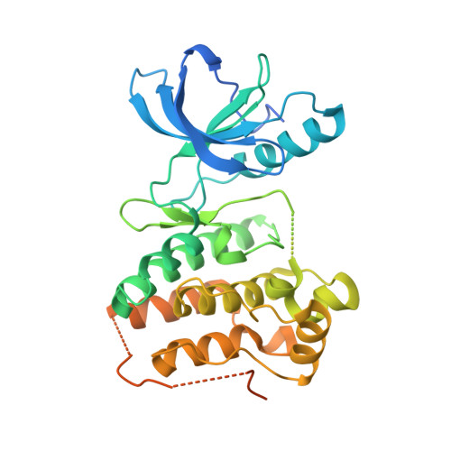
    4GK2, 4GK3, 4GK4

  • PubMed Abstract: 

    Inhibition of the tyrosine kinase erythropoietin-producing human hepatocellular carcinoma receptor B4 (EphB4) is an effective strategy for the treatment of solid tumors. We have previously reported a low nanomolar ATP-competitive inhibitor of EphB4 discovered in silico by fragment-based high-throughput docking combined with explicit solvent molecular dynamics simulations. Here we present a second generation of EphB4 inhibitors that show high inhibitory potency in both enzymatic and cell-based assays while preserving the appealing selectivity profile exhibited by the parent compound. In addition, respectable levels of antiproliferative activity for these compounds have been obtained. Finally, the binding mode predicted by docking and molecular dynamics simulations is validated by solving the crystal structures of three members of this chemical class in complex with the EphA3 tyrosine kinase whose ATP-binding site is essentially identical to that of EphB4.


  • Organizational Affiliation
    • Department of Organic Chemistry, University of Zurich, Winterthurerstrasse 190, CH-8057 Zurich, Switzerland.

Macromolecule Content 

  • Total Structure Weight: 40.88 kDa 
  • Atom Count: 2,239 
  • Modeled Residue Count: 265 
  • Deposited Residue Count: 361 
  • Unique protein chains: 1

Macromolecules

Find similar proteins by:|  3D Structure
Entity ID: 1
MoleculeChains  Sequence LengthOrganismDetailsImage
EPH receptor A3361Homo sapiensMutation(s): 0 
Gene Names: EPHA3
EC: 2.7.10.1
UniProt & NIH Common Fund Data Resources
Find proteins for P29320 (Homo sapiens)
Explore P29320 
Go to UniProtKB:  P29320
PHAROS:  P29320
GTEx:  ENSG00000044524 
Entity Groups
Sequence Clusters30% Identity50% Identity70% Identity90% Identity95% Identity100% Identity
UniProt GroupP29320
Sequence Annotations
Expand
Reference Sequence

Small Molecules

Ligands 1 Unique
IDChains Name / Formula / InChI Key2D Diagram3D Interactions
L66

Query on L66


Download:Ideal Coordinates CCD File
B [auth A]7-(5-hydroxy-2-methylphenyl)-8-(2-methoxyphenyl)-1-methyl-1H-imidazo[2,1-f]purine-2,4(3H,8H)-dione
C22 H19 N5 O4
JTWMOWRMSZZHDR-UHFFFAOYSA-N

Experimental Data & Validation

Experimental Data

  • Method: X-RAY DIFFRACTION
  • Resolution: 2.19 Å
  • R-Value Free:  0.236 (Depositor), 0.231 (DCC) 
  • R-Value Work:  0.191 (Depositor), 0.188 (DCC) 
  • R-Value Observed: 0.193 (Depositor) 
Space Group: P 1 21 1
Unit Cell:
Length ( Å )Angle ( ˚ )
a = 52.601α = 90
b = 38.195β = 101.71
c = 75.704γ = 90
Software Package:
Software NamePurpose
XDSdata scaling
PHASERphasing
PHENIXrefinement
XDSdata reduction
SCALAdata scaling

Structure Validation

View Full Validation Report


Ligand Structure Quality Assessment 


Entry History 

Deposition Data

Revision History  (Full details and data files)

  • Version 1.0: 2013-01-23
    Type: Initial release
  • Version 1.1: 2018-06-20
    Changes: Data collection
  • Version 1.2: 2023-11-08
    Changes: Data collection, Database references, Derived calculations, Refinement description