4GIE | pdb_00004gie

Crystal structure of prostaglandin F synthase from Trypanosoma cruzi bound to NADP


Experimental Data Snapshot

  • Method: X-RAY DIFFRACTION
  • Resolution: 1.25 Å
  • R-Value Free: 
    0.136 (Depositor), 0.140 (DCC) 
  • R-Value Work: 
    0.119 (Depositor) 
  • R-Value Observed: 
    0.120 (Depositor) 

Starting Model: experimental
View more details

wwPDB Validation 3D Report Full Report

Validation slider image for 4GIE

Ligand Structure Quality Assessment 


This is version 1.3 of the entry. See complete history

Literature

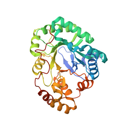
Structures of prostaglandin F synthase from the protozoa Leishmania major and Trypanosoma cruzi with NADP.

Moen, S.O.Fairman, J.W.Barnes, S.R.Sullivan, A.Nakazawa-Hewitt, S.Van Voorhis, W.C.Staker, B.L.Lorimer, D.D.Myler, P.J.Edwards, T.E.

(2015) Acta Crystallogr Sect F Struct Biol Cryst Commun 71: 609-614

  • DOI: https://doi.org/10.1107/S2053230X15006883
  • Primary Citation Related Structures: 
    4F40, 4FZI, 4G5D, 4GIE

  • PubMed Abstract: 

    The crystal structures of prostaglandin F synthase (PGF) from both Leishmania major and Trypanosoma cruzi with and without their cofactor NADP have been determined to resolutions of 2.6 Å for T. cruzi PGF, 1.25 Å for T. cruzi PGF with NADP, 1.6 Å for L. major PGF and 1.8 Å for L. major PGF with NADP. These structures were determined by molecular replacement to a final R factor of less than 18.6% (Rfree of less than 22.9%). PGF in the infectious protozoa L. major and T. cruzi is a potential therapeutic target.


  • Organizational Affiliation
    • Seattle Structural Genomics Center for Infectious Disease, USA.

Macromolecule Content 

  • Total Structure Weight: 34.39 kDa 
  • Atom Count: 2,854 
  • Modeled Residue Count: 288 
  • Deposited Residue Count: 290 
  • Unique protein chains: 1

Macromolecules

Find similar proteins by:|  3D Structure
Entity ID: 1
MoleculeChains  Sequence LengthOrganismDetailsImage
Prostaglandin F synthase290Trypanosoma cruzi strain CL BrenerMutation(s): 0 
Gene Names: Tc00.1047053511287.49
EC: 1.1.1.188 (PDB Primary Data), 1.1.1 (UniProt)
UniProt
Find proteins for Q4DJ07 (Trypanosoma cruzi (strain CL Brener))
Explore Q4DJ07 
Go to UniProtKB:  Q4DJ07
Entity Groups
Sequence Clusters30% Identity50% Identity70% Identity90% Identity95% Identity100% Identity
UniProt GroupQ4DJ07
Sequence Annotations
Expand
Reference Sequence

Small Molecules

Ligands 2 Unique
IDChains Name / Formula / InChI Key2D Diagram3D Interactions
NAP

Query on NAP


Download:Ideal Coordinates CCD File
C [auth A]NADP NICOTINAMIDE-ADENINE-DINUCLEOTIDE PHOSPHATE
C21 H28 N7 O17 P3
XJLXINKUBYWONI-NNYOXOHSSA-N
ACT

Query on ACT


Download:Ideal Coordinates CCD File
B [auth A]ACETATE ION
C2 H3 O2
QTBSBXVTEAMEQO-UHFFFAOYSA-M

Experimental Data & Validation

Experimental Data

  • Method: X-RAY DIFFRACTION
  • Resolution: 1.25 Å
  • R-Value Free:  0.136 (Depositor), 0.140 (DCC) 
  • R-Value Work:  0.119 (Depositor) 
  • R-Value Observed: 0.120 (Depositor) 
Space Group: C 1 2 1
Unit Cell:
Length ( Å )Angle ( ˚ )
a = 155.54α = 90
b = 50.34β = 94.23
c = 37.6γ = 90
Software Package:
Software NamePurpose
XSCALEdata scaling
PHASERphasing
REFMACrefinement
PDB_EXTRACTdata extraction
XDSdata reduction

Structure Validation

View Full Validation Report


Ligand Structure Quality Assessment 


Entry History 

Deposition Data

Revision History  (Full details and data files)

  • Version 1.0: 2012-08-15
    Type: Initial release
  • Version 1.1: 2015-05-13
    Changes: Database references
  • Version 1.2: 2015-06-03
    Changes: Database references
  • Version 1.3: 2023-09-13
    Changes: Data collection, Database references, Derived calculations, Refinement description