4GGM | pdb_00004ggm

Structure of LpxI


Experimental Data Snapshot

  • Method: X-RAY DIFFRACTION
  • Resolution: 2.90 Å
  • R-Value Free: 
    0.272 (Depositor), 0.272 (DCC) 
  • R-Value Work: 
    0.216 (Depositor), 0.213 (DCC) 
  • R-Value Observed: 
    0.219 (Depositor) 

wwPDB Validation 3D Report Full Report

Validation slider image for 4GGM

Ligand Structure Quality Assessment 


This is version 1.5 of the entry. See complete history

Literature

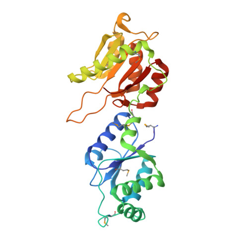
LpxI structures reveal how a lipid A precursor is synthesized.

Metzger, L.E.Lee, J.K.Finer-Moore, J.S.Raetz, C.R.Stroud, R.M.

(2012) Nat Struct Mol Biol 19: 1132-1138

  • DOI: https://doi.org/10.1038/nsmb.2393
  • Primary Citation Related Structures: 
    4GGM, 4J6E

  • PubMed Abstract: 

    Enzymes in lipid metabolism acquire and deliver hydrophobic substrates and products from within lipid bilayers. The structure at 2.55 Å of one isozyme of a constitutive enzyme in lipid A biosynthesis, LpxI from Caulobacter crescentus, has a novel fold. Two domains close around a completely sequestered substrate, UDP-2,3-diacylglucosamine, and open to release products either to the neighboring enzyme in a putative multienzyme complex or to the bilayer. Mutation analysis identifies Asp225 as key to Mg(2+)-catalyzed diphosphate hydrolysis. These structures provide snapshots of the enzymatic synthesis of a critical lipid A precursor.


  • Organizational Affiliation
    • Department of Biochemistry and Biophysics, The University of California San Francisco, San Francisco, California, USA. metzger@msg.ucsf.edu

Macromolecule Content 

  • Total Structure Weight: 31.11 kDa 
  • Atom Count: 2,148 
  • Modeled Residue Count: 277 
  • Deposited Residue Count: 283 
  • Unique protein chains: 1

Macromolecules

Find similar proteins by:|  3D Structure
Entity ID: 1
MoleculeChains  Sequence LengthOrganismDetailsImage
UDP-2,3-diacylglucosamine pyrophosphatase LpxIA [auth X]283Caulobacter vibrioides NA1000Mutation(s): 0 
Gene Names: 7330127CCNA_01987lpxI
EC: 3.6.1.54
UniProt
Find proteins for A0A0H3C8Q1 (Caulobacter vibrioides (strain NA1000 / CB15N))
Explore A0A0H3C8Q1 
Go to UniProtKB:  A0A0H3C8Q1
Entity Groups
Sequence Clusters30% Identity50% Identity70% Identity90% Identity95% Identity100% Identity
UniProt GroupA0A0H3C8Q1
Sequence Annotations
Expand
Reference Sequence

Small Molecules

Ligands 2 Unique
IDChains Name / Formula / InChI Key2D Diagram3D Interactions
LP5

Query on LP5


Download:Ideal Coordinates CCD File
C [auth X](R)-((2R,3S,4R,5R,6R)-3-HYDROXY-2-(HYDROXYMETHYL)-5-((R)-3-HYDROXYTETRADECANAMIDO)-6-(PHOSPHONOOXY)TETRAHYDRO-2H-PYRAN-4-YL) 3-HYDROXYTETRADECANOATE
C34 H66 N O12 P
HEHQDWUWJVPREQ-XQJZMFRCSA-N
MG

Query on MG


Download:Ideal Coordinates CCD File
B [auth X]MAGNESIUM ION
Mg
JLVVSXFLKOJNIY-UHFFFAOYSA-N
Modified Residues  1 Unique
IDChains TypeFormula2D DiagramParent
MSE
Query on MSE
A [auth X]L-PEPTIDE LINKINGC5 H11 N O2 SeMET

Experimental Data & Validation

Experimental Data

  • Method: X-RAY DIFFRACTION
  • Resolution: 2.90 Å
  • R-Value Free:  0.272 (Depositor), 0.272 (DCC) 
  • R-Value Work:  0.216 (Depositor), 0.213 (DCC) 
  • R-Value Observed: 0.219 (Depositor) 
Space Group: I 41 2 2
Unit Cell:
Length ( Å )Angle ( ˚ )
a = 74.286α = 90
b = 74.286β = 90
c = 364.644γ = 90
Software Package:
Software NamePurpose
Blu-Icedata collection
PHASERphasing
PHENIXrefinement
MOSFLMdata reduction
SCALAdata scaling

Structure Validation

View Full Validation Report


Ligand Structure Quality Assessment 


Entry History 

Revision History  (Full details and data files)

  • Version 1.0: 2012-10-03
    Type: Initial release
  • Version 1.1: 2012-11-14
    Changes: Database references
  • Version 1.2: 2012-11-21
    Changes: Database references
  • Version 1.3: 2013-04-17
    Changes: Structure summary
  • Version 1.4: 2017-10-25
    Changes: Author supporting evidence
  • Version 1.5: 2024-11-06
    Changes: Data collection, Database references, Derived calculations, Structure summary