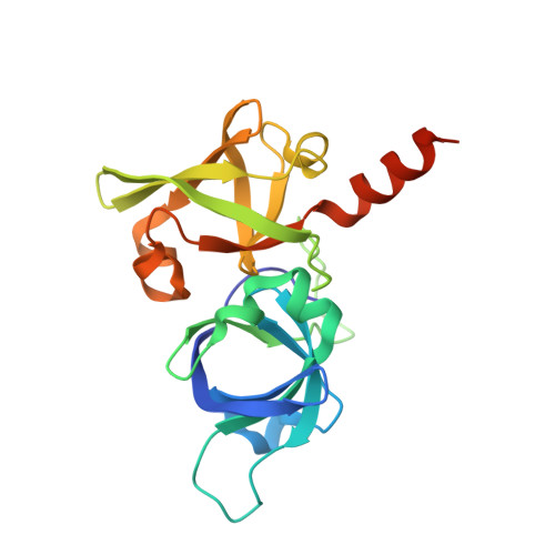
Crystallographic and kinetic study of riboflavin synthase from Brucella abortus, a chemotherapeutic target with an enhanced intrinsic flexibility.
Serer, M.I., Bonomi, H.R., Guimaraes, B.G., Rossi, R.C., Goldbaum, F.A., Klinke, S.(2014) Acta Crystallogr D Biol Crystallogr 70: 1419-1434
- PubMed: 24816110
- DOI: https://doi.org/10.1107/S1399004714005161
- Primary Citation of Related Structures:
4E0F, 4FXU, 4G6I, 4GQN - PubMed Abstract:
Riboflavin synthase (RS) catalyzes the last step of riboflavin biosynthesis in microorganisms and plants, which corresponds to the dismutation of two molecules of 6,7-dimethyl-8-ribityllumazine to yield one molecule of riboflavin and one molecule of 5-amino-6-ribitylamino-2,4(1H,3H)-pyrimidinedione. Owing to the absence of this enzyme in animals and the fact that most pathogenic bacteria show a strict dependence on riboflavin biosynthesis, RS has been proposed as a potential target for antimicrobial drug development. Eubacterial, fungal and plant RSs assemble as homotrimers lacking C3 symmetry. Each monomer can bind two substrate molecules, yet there is only one active site for the whole enzyme, which is located at the interface between two neighbouring chains. This work reports the crystallographic structure of RS from the pathogenic bacterium Brucella abortus (the aetiological agent of the disease brucellosis) in its apo form, in complex with riboflavin and in complex with two different product analogues, being the first time that the structure of an intact RS trimer with bound ligands has been solved. These crystal models support the hypothesis of enhanced flexibility in the particle and also highlight the role of the ligands in assembling the unique active site. Kinetic and binding studies were also performed to complement these findings. The structural and biochemical information generated may be useful for the rational design of novel RS inhibitors with antimicrobial activity.
- Fundación Instituto Leloir, IIBBA-CONICET, Avenida Patricias Argentinas 435, C1405BWE Buenos Aires, Argentina.
Organizational Affiliation:


