4G2G | pdb_00004g2g

Crystal structure of Mycobacterium tuberculosis CYP121 in complex with 4,4'-(1H-1,2,3-triazole-1,5-diyl)diphenol


Experimental Data Snapshot

  • Method: X-RAY DIFFRACTION
  • Resolution: 2.25 Å
  • R-Value Free: 
    0.217 (Depositor), 0.214 (DCC) 
  • R-Value Work: 
    0.148 (Depositor), 0.147 (DCC) 
  • R-Value Observed: 
    0.151 (Depositor) 

Starting Model: experimental
View more details

wwPDB Validation 3D Report Full Report

Validation slider image for 4G2G

Ligand Structure Quality Assessment 


This is version 1.2 of the entry. See complete history

Literature

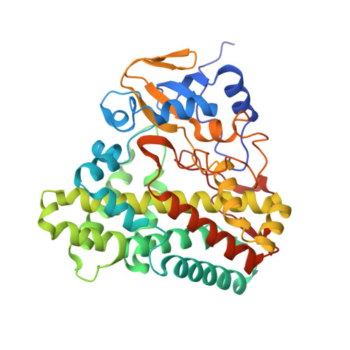
Application of Fragment Screening and Merging to the Discovery of Inhibitors of the Mycobacterium tuberculosis Cytochrome P450 CYP121

Hudson, S.A.McLean, K.J.Surade, S.Yang, Y.-Q.Leys, D.Ciulli, A.Munro, A.W.Abell, C.

(2012) Angew Chem Int Ed Engl 51: 9311-9316

Macromolecule Content 

  • Total Structure Weight: 44.58 kDa 
  • Atom Count: 3,570 
  • Modeled Residue Count: 395 
  • Deposited Residue Count: 395 
  • Unique protein chains: 1

Macromolecules

Find similar proteins by:|  3D Structure
Entity ID: 1
MoleculeChains  Sequence LengthOrganismDetailsImage
Cytochrome P450 121395Mycobacterium tuberculosisMutation(s): 0 
Gene Names: Rv2276
EC: 1.14 (PDB Primary Data), 1.14.19.70 (UniProt)
UniProt
Find proteins for P9WPP7 (Mycobacterium tuberculosis (strain ATCC 25618 / H37Rv))
Explore P9WPP7 
Go to UniProtKB:  P9WPP7
Entity Groups
Sequence Clusters30% Identity50% Identity70% Identity90% Identity95% Identity100% Identity
UniProt GroupP9WPP7
Sequence Annotations
Expand
Reference Sequence

Small Molecules

Ligands 4 Unique
IDChains Name / Formula / InChI Key2D Diagram3D Interactions
HEM

Query on HEM


Download:Ideal Coordinates CCD File
B [auth A]PROTOPORPHYRIN IX CONTAINING FE
C34 H32 Fe N4 O4
KABFMIBPWCXCRK-RGGAHWMASA-L
TDH

Query on TDH


Download:Ideal Coordinates CCD File
I [auth A]4,4'-(1H-1,2,3-triazole-1,5-diyl)diphenol
C14 H11 N3 O2
VJAKQEJLSICLIC-UHFFFAOYSA-N
SO4

Query on SO4


Download:Ideal Coordinates CCD File
C [auth A],
D [auth A],
E [auth A],
F [auth A]
SULFATE ION
O4 S
QAOWNCQODCNURD-UHFFFAOYSA-L
DMS

Query on DMS


Download:Ideal Coordinates CCD File
G [auth A],
H [auth A]
DIMETHYL SULFOXIDE
C2 H6 O S
IAZDPXIOMUYVGZ-UHFFFAOYSA-N

Experimental Data & Validation

Experimental Data

  • Method: X-RAY DIFFRACTION
  • Resolution: 2.25 Å
  • R-Value Free:  0.217 (Depositor), 0.214 (DCC) 
  • R-Value Work:  0.148 (Depositor), 0.147 (DCC) 
  • R-Value Observed: 0.151 (Depositor) 
Space Group: P 65 2 2
Unit Cell:
Length ( Å )Angle ( ˚ )
a = 77.365α = 90
b = 77.365β = 90
c = 263.645γ = 120
Software Package:
Software NamePurpose
PROTEUM PLUSdata collection
REFMACrefinement
PROTEUM PLUSdata reduction
PROTEUM PLUSdata scaling
REFMACphasing

Structure Validation

View Full Validation Report


Ligand Structure Quality Assessment 


Entry History 

Deposition Data

  • Released Date: 2012-09-05 
  • Deposition Author(s): Hudson, S.A.

Revision History  (Full details and data files)

  • Version 1.0: 2012-09-05
    Type: Initial release
  • Version 1.1: 2012-09-19
    Changes: Database references
  • Version 1.2: 2023-11-08
    Changes: Data collection, Database references, Derived calculations, Refinement description