4G1U

X-ray structure of the bacterial heme transporter HmuUV from Yersinia pestis


Experimental Data Snapshot

  • Method: X-RAY DIFFRACTION
  • Resolution: 3.01 Å
  • R-Value Free: 0.295 
  • R-Value Work: 0.263 
  • R-Value Observed: 0.265 

wwPDB Validation   3D Report Full Report


This is version 1.1 of the entry. See complete history


Literature

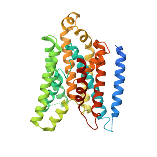
X-ray structure of the Yersinia pestis heme transporter HmuUV.

Woo, J.-S.Zeltina, A.Goetz, B.A.Locher, K.P.

(2012) Nat Struct Mol Biol 19: 1310-1315

  • DOI: https://doi.org/10.1038/nsmb.2417
  • Primary Citation of Related Structures:  
    4G1U

  • PubMed Abstract: 

    HmuUV is a bacterial ATP-binding cassette (ABC) transporter that catalyzes heme uptake into the cytoplasm of the gram-negative pathogen Yersinia pestis. We report the crystal structure of HmuUV at 3.0 Å resolution in a nucleotide-free state, which features a heme translocation pathway in an outward-facing conformation, poised to accept a heme from the cognate periplasmic binding protein HmuT. A new assay allowed us to determine in vitro rates of HmuUV-catalyzed heme transport into proteoliposomes and to establish the role of conserved residues in the translocation pathway of HmuUV and at the interface with HmuT. Differences in architecture relative to the related vitamin B(12) transporter BtuCD suggest an adaptation of HmuUV for its smaller substrate. Our study also suggests that type II ABC importers, which include bacterial iron-siderophore, heme and cobalamin transporters, have a coupling mechanism distinct from that of other ABC transporters.


  • Organizational Affiliation

    Institute of Molecular Biology and Biophysics, Eidgenössische Technische Hochschule (ETH) Zürich, CH-8093 Zürich, Switzerland.


Macromolecules
Find similar proteins by:  (by identity cutoff)  |  3D Structure
Entity ID: 1
MoleculeChains Sequence LengthOrganismDetailsImage
Hemin transport system permease protein hmuU
A, B
357Yersinia pestisMutation(s): 0 
Gene Names: hmuUYPO0280y0540YP_0435
Membrane Entity: Yes 
UniProt
Find proteins for Q56992 (Yersinia pestis)
Explore Q56992 
Go to UniProtKB:  Q56992
Entity Groups  
Sequence Clusters30% Identity50% Identity70% Identity90% Identity95% Identity100% Identity
UniProt GroupQ56992
Sequence Annotations
Expand
  • Reference Sequence
Find similar proteins by:  (by identity cutoff)  |  3D Structure
Entity ID: 2
MoleculeChains Sequence LengthOrganismDetailsImage
Hemin import ATP-binding protein HmuV
C, D
266Yersinia pestisMutation(s): 0 
Gene Names: hmuVYPO0279y0539YP_0434
EC: 3.6.3
Membrane Entity: Yes 
UniProt
Find proteins for Q56993 (Yersinia pestis)
Explore Q56993 
Go to UniProtKB:  Q56993
Entity Groups  
Sequence Clusters30% Identity50% Identity70% Identity90% Identity95% Identity100% Identity
UniProt GroupQ56993
Sequence Annotations
Expand
  • Reference Sequence
Experimental Data & Validation

Experimental Data

  • Method: X-RAY DIFFRACTION
  • Resolution: 3.01 Å
  • R-Value Free: 0.295 
  • R-Value Work: 0.263 
  • R-Value Observed: 0.265 
  • Space Group: P 1 21 1
Unit Cell:
Length ( Å )Angle ( ˚ )
a = 100.87α = 90
b = 101.23β = 91.3
c = 105.21γ = 90
Software Package:
Software NamePurpose
XSCALEdata scaling
PHENIXrefinement
PDB_EXTRACTdata extraction
XDSdata scaling
SHELXSphasing

Structure Validation

View Full Validation Report


Entry History 

Deposition Data

Revision History  (Full details and data files)

  • Version 1.0: 2012-12-19
    Type: Initial release
  • Version 1.1: 2024-03-20
    Changes: Data collection, Database references, Derived calculations