4G1H

Group B Streptococcus Pilus Island 1 Sortase C2


Experimental Data Snapshot

  • Method: X-RAY DIFFRACTION
  • Resolution: 1.80 Å
  • R-Value Free: 0.251 
  • R-Value Work: 0.212 
  • R-Value Observed: 0.216 

wwPDB Validation   3D Report Full Report


This is version 1.1 of the entry. See complete history


Literature

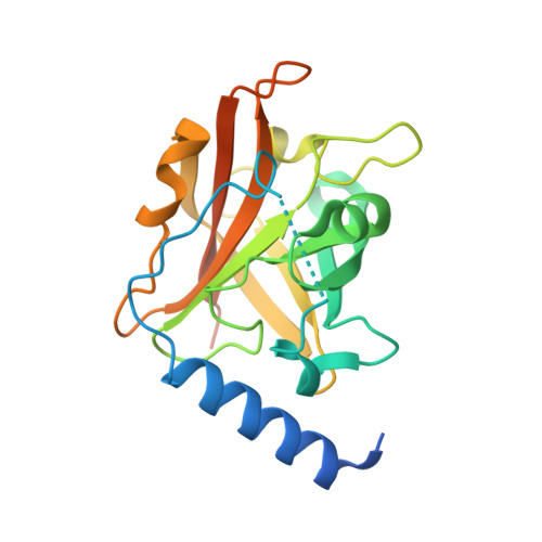
Structural basis for group B streptococcus pilus 1 sortases C regulation and specificity.

Cozzi, R.Prigozhin, D.Rosini, R.Abate, F.Bottomley, M.J.Grandi, G.Telford, J.L.Rinaudo, C.D.Maione, D.Alber, T.

(2012) PLoS One 7: e49048-e49048

  • DOI: https://doi.org/10.1371/journal.pone.0049048
  • Primary Citation of Related Structures:  
    4G1H, 4G1J

  • PubMed Abstract: 

    Gram-positive bacteria assemble pili through class C sortase enzymes specialized in polymerizing pilin subunits into covalently linked, high-molecular-weight, elongated structures. Here we report the crystal structures of two class C sortases (SrtC1 and SrtC2) from Group B Streptococcus (GBS) Pilus Island 1. The structures show that both sortases are comprised of two domains: an 8-stranded β-barrel catalytic core conserved among all sortase family members and a flexible N-terminal region made of two α-helices followed by a loop, known as the lid, which acts as a pseudo-substrate. In vitro experiments performed with recombinant SrtC enzymes lacking the N-terminal portion demonstrate that this region of the enzyme is dispensable for catalysis but may have key roles in substrate specificity and regulation. Moreover, in vitro FRET-based assays show that the LPXTG motif common to many sortase substrates is not the sole determinant of sortase C specificity during pilin protein recognition.


  • Organizational Affiliation

    Novartis Vaccines and Diagnostics, Siena, Italy.


Macromolecules
Find similar proteins by:  (by identity cutoff)  |  3D Structure
Entity ID: 1
MoleculeChains Sequence LengthOrganismDetailsImage
Sortase family protein215Streptococcus agalactiae serogroup VMutation(s): 0 
Gene Names: SAG0648
UniProt
Find proteins for Q8E0S6 (Streptococcus agalactiae serotype V (strain ATCC BAA-611 / 2603 V/R))
Explore Q8E0S6 
Go to UniProtKB:  Q8E0S6
Entity Groups  
Sequence Clusters30% Identity50% Identity70% Identity90% Identity95% Identity100% Identity
UniProt GroupQ8E0S6
Sequence Annotations
Expand
  • Reference Sequence
Small Molecules
Ligands 1 Unique
IDChains Name / Formula / InChI Key2D Diagram3D Interactions
CA
Query on CA

Download Ideal Coordinates CCD File 
B [auth A]CALCIUM ION
Ca
BHPQYMZQTOCNFJ-UHFFFAOYSA-N
Experimental Data & Validation

Experimental Data

  • Method: X-RAY DIFFRACTION
  • Resolution: 1.80 Å
  • R-Value Free: 0.251 
  • R-Value Work: 0.212 
  • R-Value Observed: 0.216 
  • Space Group: P 41 21 2
Unit Cell:
Length ( Å )Angle ( ˚ )
a = 60.435α = 90
b = 60.435β = 90
c = 102.242γ = 90
Software Package:
Software NamePurpose
PHENIXrefinement
HKL-2000data reduction
HKL-2000data scaling

Structure Validation

View Full Validation Report


Entry History 

Deposition Data

Revision History  (Full details and data files)

  • Version 1.0: 2012-12-05
    Type: Initial release
  • Version 1.1: 2024-02-28
    Changes: Data collection, Database references, Derived calculations