4FXX

Structure of SF1 coiled-coil domain


Experimental Data Snapshot

  • Method: X-RAY DIFFRACTION
  • Resolution: 2.48 Å
  • R-Value Free: 0.270 
  • R-Value Work: 0.236 
  • R-Value Observed: 0.239 

wwPDB Validation   3D Report Full Report


This is version 1.2 of the entry. See complete history


Literature

Structure of Phosphorylated SF1 Bound to U2AF(65) in an Essential Splicing Factor Complex.

Wang, W.Maucuer, A.Gupta, A.Manceau, V.Thickman, K.R.Bauer, W.J.Kennedy, S.D.Wedekind, J.E.Green, M.R.Kielkopf, C.L.

(2013) Structure 21: 197-208

  • DOI: https://doi.org/10.1016/j.str.2012.10.020
  • Primary Citation of Related Structures:  
    4FXW, 4FXX

  • PubMed Abstract: 

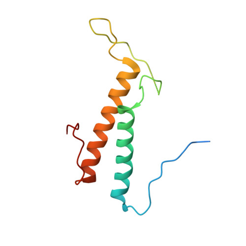
    The essential splicing factors U2AF⁶⁵ and SF1 cooperatively bind consensus sequences at the 3' end of introns. Phosphorylation of SF1 on a highly conserved "SPSP" motif enhances its interaction with U2AF⁶⁵ and the pre-mRNA. Here, we reveal that phosphorylation induces essential conformational changes in SF1 and in the SF1/U2AF⁶⁵/3' splice site complex. Crystal structures of the phosphorylated (P)SF1 domain bound to the C-terminal domain of U2AF⁶⁵ at 2.29 Å resolution and of the unphosphorylated SF1 domain at 2.48 Å resolution demonstrate that phosphorylation induces a disorder-to-order transition within a previously unknown SF1/U2AF⁶⁵ interface. We find by small-angle X-ray scattering that the local folding of the SPSP motif transduces into global conformational changes in the nearly full-length (P)SF1/U2AF⁶⁵/3' splice site assembly. We further determine that SPSP phosphorylation and the SF1/U2AF⁶⁵ interface are essential in vivo. These results offer a structural prototype for phosphorylation-dependent control of pre-mRNA splicing factors.


  • Organizational Affiliation

    Center for RNA Biology and Department of Biochemistry and Biophysics, University of Rochester School of Medicine and Dentistry, Rochester, NY 14642, USA.


Macromolecules
Find similar proteins by:  (by identity cutoff)  |  3D Structure
Entity ID: 1
MoleculeChains Sequence LengthOrganismDetailsImage
Splicing factor 1
A, B, C, D
112Homo sapiensMutation(s): 0 
Gene Names: SF1ZFM1ZNF162
UniProt & NIH Common Fund Data Resources
Find proteins for Q15637 (Homo sapiens)
Explore Q15637 
Go to UniProtKB:  Q15637
PHAROS:  Q15637
GTEx:  ENSG00000168066 
Entity Groups  
Sequence Clusters30% Identity50% Identity70% Identity90% Identity95% Identity100% Identity
UniProt GroupQ15637
Sequence Annotations
Expand
  • Reference Sequence
Small Molecules
Ligands 2 Unique
IDChains Name / Formula / InChI Key2D Diagram3D Interactions
MLI
Query on MLI

Download Ideal Coordinates CCD File 
F [auth A],
K [auth C]
MALONATE ION
C3 H2 O4
OFOBLEOULBTSOW-UHFFFAOYSA-L
IMD
Query on IMD

Download Ideal Coordinates CCD File 
E [auth A]
G [auth B]
H [auth B]
I [auth C]
J [auth C]
E [auth A],
G [auth B],
H [auth B],
I [auth C],
J [auth C],
L [auth D],
M [auth D],
N [auth D]
IMIDAZOLE
C3 H5 N2
RAXXELZNTBOGNW-UHFFFAOYSA-O
Experimental Data & Validation

Experimental Data

  • Method: X-RAY DIFFRACTION
  • Resolution: 2.48 Å
  • R-Value Free: 0.270 
  • R-Value Work: 0.236 
  • R-Value Observed: 0.239 
  • Space Group: C 1 2 1
Unit Cell:
Length ( Å )Angle ( ˚ )
a = 96.03α = 90
b = 37.97β = 107.38
c = 144.68γ = 90
Software Package:
Software NamePurpose
SCALAdata scaling
PHENIXrefinement
PDB_EXTRACTdata extraction
Web-Icedata collection
MOSFLMdata reduction
PHENIXphasing

Structure Validation

View Full Validation Report


Entry History 

Deposition Data

Revision History  (Full details and data files)

  • Version 1.0: 2013-01-16
    Type: Initial release
  • Version 1.1: 2013-02-27
    Changes: Database references
  • Version 1.2: 2024-02-28
    Changes: Data collection, Database references, Derived calculations, Refinement description