


Crystal Structure of the ERK2 complexed with EK3
Kang, Y.N., Stuckey, J.A., Xie, X.To be published.
Experimental Data Snapshot
Entity ID: 1 | |||||
|---|---|---|---|---|---|
| Molecule | Chains | Sequence Length | Organism | Details | Image |
| Mitogen-activated protein kinase 1 | 360 | Homo sapiens | Mutation(s): 0 Gene Names: ERK2, MAPK1, PRKM1, PRKM2 EC: 2.7.11.24 | 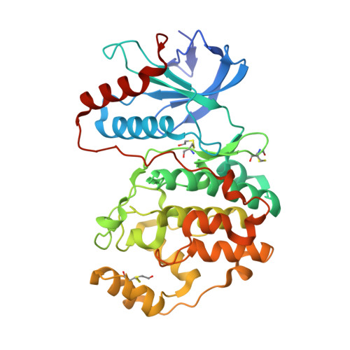 | |
UniProt & NIH Common Fund Data Resources | |||||
PHAROS: P28482 GTEx: ENSG00000100030 | |||||
Entity Groups | |||||
| Sequence Clusters | 30% Identity50% Identity70% Identity90% Identity95% Identity100% Identity | ||||
| UniProt Group | P28482 | ||||
Sequence AnnotationsExpand | |||||
Reference Sequence | |||||
| Ligands 3 Unique | |||||
|---|---|---|---|---|---|
| ID | Chains | Name / Formula / InChI Key | 2D Diagram | 3D Interactions | |
| EK3 Download:Ideal Coordinates CCD File | B [auth A] | N-cyclohexyl-4-[3-(4-fluorophenyl)-1H-pyrazol-4-yl]pyridin-2-amine C20 H21 F N4 JMLIPRNMUGOAAS-UHFFFAOYSA-N |  | ||
| GOL Download:Ideal Coordinates CCD File | K [auth A], L [auth A] | GLYCEROL C3 H8 O3 PEDCQBHIVMGVHV-UHFFFAOYSA-N |  | ||
| EDO Download:Ideal Coordinates CCD File | C [auth A] D [auth A] E [auth A] F [auth A] G [auth A] | 1,2-ETHANEDIOL C2 H6 O2 LYCAIKOWRPUZTN-UHFFFAOYSA-N |  | ||
| Modified Residues 1 Unique | |||||
|---|---|---|---|---|---|
| ID | Chains | Type | Formula | 2D Diagram | Parent |
| CME Query on CME | A | L-PEPTIDE LINKING | C5 H11 N O3 S2 |  | CYS |
| Length ( Å ) | Angle ( ˚ ) |
|---|---|
| a = 48.8 | α = 90 |
| b = 69.92 | β = 108.68 |
| c = 59.95 | γ = 90 |
| Software Name | Purpose |
|---|---|
| SCALEPACK | data scaling |
| BUSTER-TNT | refinement |
| PDB_EXTRACT | data extraction |
| BUSTER | refinement |


