


Crystal structure of methyltransferase domain of human RNA (guanine-9-) methyltransferase domain containing protein 2
Zeng, H., Dong, A., Loppnau, P., Tempel, W., Bountra, C., Weigelt, J., Arrowsmith, C.H., Edwards, A.M., Wu, H.To be published.
Experimental Data Snapshot
Entity ID: 1 | |||||
|---|---|---|---|---|---|
| Molecule | Chains | Sequence Length | Organism | Details | Image |
| RNA (guanine-9-)-methyltransferase domain-containing protein 2 | 197 | Homo sapiens | Mutation(s): 0 Gene Names: RG9MTD2 EC: 2.1.1 (PDB Primary Data), 2.1.1.221 (UniProt) | 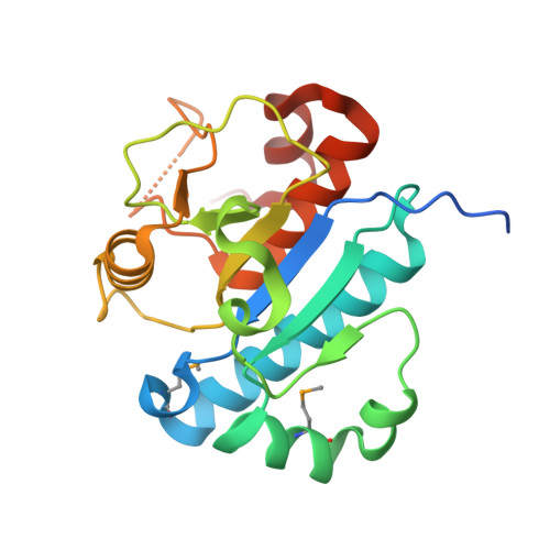 | |
UniProt & NIH Common Fund Data Resources | |||||
PHAROS: Q8TBZ6 GTEx: ENSG00000145331 | |||||
Entity Groups | |||||
| Sequence Clusters | 30% Identity50% Identity70% Identity90% Identity95% Identity100% Identity | ||||
| UniProt Group | Q8TBZ6 | ||||
Sequence AnnotationsExpand | |||||
Reference Sequence | |||||
| Ligands 4 Unique | |||||
|---|---|---|---|---|---|
| ID | Chains | Name / Formula / InChI Key | 2D Diagram | 3D Interactions | |
| SAH Download:Ideal Coordinates CCD File | C [auth A], O [auth B] | S-ADENOSYL-L-HOMOCYSTEINE C14 H20 N6 O5 S ZJUKTBDSGOFHSH-WFMPWKQPSA-N |  | ||
| EDO Download:Ideal Coordinates CCD File | D [auth A], P [auth B], Q [auth B] | 1,2-ETHANEDIOL C2 H6 O2 LYCAIKOWRPUZTN-UHFFFAOYSA-N |  | ||
| K Download:Ideal Coordinates CCD File | R [auth B] | POTASSIUM ION K NPYPAHLBTDXSSS-UHFFFAOYSA-N |  | ||
| UNX Download:Ideal Coordinates CCD File | AA [auth B] BA [auth B] E [auth A] F [auth A] G [auth A] | UNKNOWN ATOM OR ION X |  | ||
| Modified Residues 1 Unique | |||||
|---|---|---|---|---|---|
| ID | Chains | Type | Formula | 2D Diagram | Parent |
| MSE Query on MSE | A, B | L-PEPTIDE LINKING | C5 H11 N O2 Se |  | MET |
| Length ( Å ) | Angle ( ˚ ) |
|---|---|
| a = 37.097 | α = 100.43 |
| b = 43.309 | β = 90.03 |
| c = 60.347 | γ = 92.88 |
| Software Name | Purpose |
|---|---|
| CLS | data collection |
| SOLVE | phasing |
| RESOLVE | model building |
| BUSTER | refinement |
| Coot | model building |
| HKL-2000 | data reduction |
| HKL-2000 | data scaling |
| RESOLVE | phasing |


