4FDK

F78L Tt H-NOX


Experimental Data Snapshot

  • Method: X-RAY DIFFRACTION
  • Resolution: 2.10 Å
  • R-Value Free: 0.231 
  • R-Value Work: 0.170 
  • R-Value Observed: 0.173 

wwPDB Validation   3D Report Full Report


Ligand Structure Quality Assessment 


This is version 1.3 of the entry. See complete history


Literature

Porphyrin pi-stacking in a heme protein scaffold tunes gas ligand affinity.

Weinert, E.E.Phillips-Piro, C.M.Marletta, M.A.

(2013) J Inorg Biochem 127C: 7-12

  • DOI: https://doi.org/10.1016/j.jinorgbio.2013.06.004
  • Primary Citation of Related Structures:  
    4FDK

  • PubMed Abstract: 

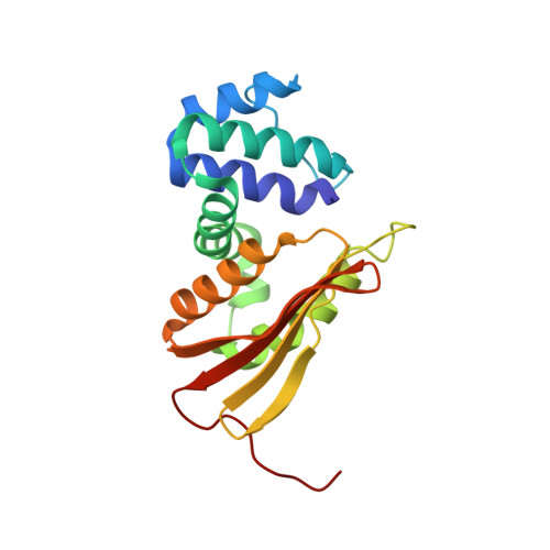
    The role of π-stacking in controlling redox and ligand binding properties of porphyrins has been of interest for many years. The recent discovery of H-NOX domains has provided a model system to investigate the role of porphyrin π-stacking within a heme protein scaffold. Removal of a phenylalanine-porphyrin π-stack dramatically increased O2, NO, and CO affinities and caused changes in redox potential (~40mV) without any structural changes. These results suggest that small changes in redox potential affect ligand affinity and that π-stacking may provide a novel route to engineer heme protein properties for new functions.


  • Organizational Affiliation

    Department of Chemistry, Emory University, Atlanta, GA 30322, United States.


Macromolecules
Find similar proteins by:  (by identity cutoff)  |  3D Structure
Entity ID: 1
MoleculeChains Sequence LengthOrganismDetailsImage
Methyl-accepting chemotaxis protein
A, B
188Caldanaerobacter subterraneus subsp. tengcongensis MB4Mutation(s): 1 
Gene Names: Tar4TTE0680
UniProt
Find proteins for Q8RBX6 (Caldanaerobacter subterraneus subsp. tengcongensis (strain DSM 15242 / JCM 11007 / NBRC 100824 / MB4))
Explore Q8RBX6 
Go to UniProtKB:  Q8RBX6
Entity Groups  
Sequence Clusters30% Identity50% Identity70% Identity90% Identity95% Identity100% Identity
UniProt GroupQ8RBX6
Sequence Annotations
Expand
  • Reference Sequence
Experimental Data & Validation

Experimental Data

  • Method: X-RAY DIFFRACTION
  • Resolution: 2.10 Å
  • R-Value Free: 0.231 
  • R-Value Work: 0.170 
  • R-Value Observed: 0.173 
  • Space Group: P 21 21 2
Unit Cell:
Length ( Å )Angle ( ˚ )
a = 80.552α = 90
b = 127.426β = 90
c = 42.935γ = 90
Software Package:
Software NamePurpose
PHENIXrefinement
PHASERphasing
ELVESrefinement
HKL-2000data reduction
HKL-2000data scaling

Structure Validation

View Full Validation Report


Ligand Structure Quality Assessment 


Entry History 

Deposition Data

Revision History  (Full details and data files)

  • Version 1.0: 2013-08-28
    Type: Initial release
  • Version 1.1: 2017-11-15
    Changes: Refinement description
  • Version 1.2: 2019-07-17
    Changes: Data collection, Refinement description
  • Version 1.3: 2023-09-13
    Changes: Data collection, Database references, Derived calculations, Refinement description