4ESI | pdb_00004esi

Structure of ricin A chain bound with N-((1H-1,2,3-triazol-4-yl)methyl-2-amino-4-oxo-3,4-dihydropteridine-7-carboxamide


Experimental Data Snapshot

  • Method: X-RAY DIFFRACTION
  • Resolution: 1.87 Å
  • R-Value Free: 
    0.285 (Depositor), 0.283 (DCC) 
  • R-Value Work: 
    0.229 (Depositor), 0.227 (DCC) 
  • R-Value Observed: 
    0.232 (Depositor) 

Starting Model: experimental
View more details

wwPDB Validation 3D Report Full Report

Validation slider image for 4ESI

Ligand Structure Quality Assessment 


This is version 1.2 of the entry. See complete history

Literature

Optimized 5-membered heterocycle-linked pterins for the inhibition of Ricin Toxin A.

Pruet, J.M.Saito, R.Manzano, L.A.Jasheway, K.R.Wiget, P.A.Kamat, I.Anslyn, E.V.Robertus, J.D.

(2012) ACS Med Chem Lett 3: 588-591

  • DOI: https://doi.org/10.1021/ml300099t
  • Primary Citation Related Structures: 
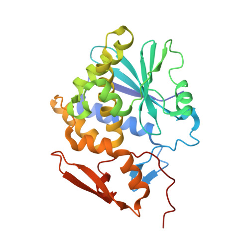
    4ESI

  • PubMed Abstract: 

    The optimization of a series of pterin amides for use as Ricin Toxin A (RTA) inhibitors is reported. Based upon crystallographic data of a previous furan-linked pterin, various expanded furans were synthesized, linked to the pterin and tested for inhibition. Concurrently, hetero-analogs of furan were explored, leading to the discovery of more potent triazol-linked pterins. Additionally, we discuss a dramatic improvement in the synthesis of these pterin amides via a dual role by diazabicycloundecene (DBU). This synthetic enhancement facilitates rapid diversification of the previously challenging pterin heterocycle, potentially aiding future medicinal research involving this structure.


  • Organizational Affiliation
    • Department of Chemistry and Biochemistry, University of Texas at Austin, 1 University Station A1590, Austin, TX 78712, USA.

Macromolecule Content 

  • Total Structure Weight: 30.22 kDa 
  • Atom Count: 2,248 
  • Modeled Residue Count: 261 
  • Deposited Residue Count: 267 
  • Unique protein chains: 1

Macromolecules

Find similar proteins by:|  3D Structure
Entity ID: 1
MoleculeChains  Sequence LengthOrganismDetailsImage
Ricin267Ricinus communisMutation(s): 0 
EC: 3.2.2.22
UniProt
Find proteins for P02879 (Ricinus communis)
Explore P02879 
Go to UniProtKB:  P02879
Entity Groups
Sequence Clusters30% Identity50% Identity70% Identity90% Identity95% Identity100% Identity
UniProt GroupP02879
Sequence Annotations
Expand
Reference Sequence

Small Molecules

Ligands 1 Unique
IDChains Name / Formula / InChI Key2D Diagram3D Interactions
0RB

Query on 0RB


Download:Ideal Coordinates CCD File
B [auth A]2-amino-4-oxo-N-(1H-1,2,3-triazol-5-ylmethyl)-1,4-dihydropteridine-7-carboxamide
C10 H9 N9 O2
IOLZYHQWMHJFBL-UHFFFAOYSA-N

Experimental Data & Validation

Experimental Data

  • Method: X-RAY DIFFRACTION
  • Resolution: 1.87 Å
  • R-Value Free:  0.285 (Depositor), 0.283 (DCC) 
  • R-Value Work:  0.229 (Depositor), 0.227 (DCC) 
  • R-Value Observed: 0.232 (Depositor) 
Space Group: P 41 21 2
Unit Cell:
Length ( Å )Angle ( ˚ )
a = 68.359α = 90
b = 68.359β = 90
c = 141.088γ = 90
Software Package:
Software NamePurpose
DENZOdata reduction
SCALEPACKdata scaling
REFMACrefinement
PDB_EXTRACTdata extraction
StructureStudiodata collection
PHASERphasing

Structure Validation

View Full Validation Report


Ligand Structure Quality Assessment 


Entry History 

Deposition Data

Revision History  (Full details and data files)

  • Version 1.0: 2012-10-31
    Type: Initial release
  • Version 1.1: 2017-11-15
    Changes: Refinement description
  • Version 1.2: 2023-09-13
    Changes: Data collection, Database references, Derived calculations, Refinement description