4EOU

Crystal structure of E. coli dihydrodipicolinate synthase with pyruvate and succinic semi-aldehyde bound in active site


Experimental Data Snapshot

  • Method: X-RAY DIFFRACTION
  • Resolution: 2.30 Å
  • R-Value Free: 0.198 
  • R-Value Work: 0.143 
  • R-Value Observed: 0.146 

wwPDB Validation   3D Report Full Report


This is version 1.4 of the entry. See complete history


Literature

The crystal structure of dihydrodipicolinate synthase from Escherichia coli with bound pyruvate and succinic acid semialdehyde: Unambiguous resolution of the stereochemistry of the condensation product.

Boughton, B.A.Dobson, R.C.Hutton, C.A.

(2012) Proteins 80: 2117-2122

  • DOI: https://doi.org/10.1002/prot.24106
  • Primary Citation of Related Structures:  
    4EOU

  • PubMed Abstract: 

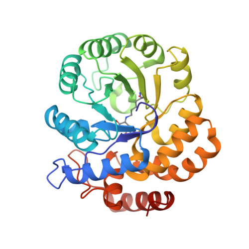
    The crystal structure of Escherichia coli dihydrodipicolinate synthase with pyruvate and substrate analogue succinic acid semialdehyde condensed with the active site lysine-161 was solved to a resolution of 2.3 Å. Comparative analysis to a previously reported structure both resolves the configuration at the aldol addition center, where the final addition product clearly displays the (S)-configuration, and the final conformation of the adduct within the active site. Direct comparison to two other crystal structures found in the Protein Data Bank, 1YXC, and 3DU0, demonstrates significant similarity between the active site residues of these structures.


  • Organizational Affiliation

    School of Chemistry, Bio21 Molecular Science and Biotechnology Institute, The University of Melbourne, Parkville, Victoria 3010, Australia. baboug@unimelb.edu.au


Macromolecules
Find similar proteins by:  (by identity cutoff)  |  3D Structure
Entity ID: 1
MoleculeChains Sequence LengthOrganismDetailsImage
Dihydrodipicolinate synthase
A, B
292Escherichia coliMutation(s): 0 
EC: 4.2.1.52
UniProt
Find proteins for P0A6L2 (Escherichia coli (strain K12))
Explore P0A6L2 
Go to UniProtKB:  P0A6L2
Entity Groups  
Sequence Clusters30% Identity50% Identity70% Identity90% Identity95% Identity100% Identity
UniProt GroupP0A6L2
Sequence Annotations
Expand
  • Reference Sequence
Experimental Data & Validation

Experimental Data

  • Method: X-RAY DIFFRACTION
  • Resolution: 2.30 Å
  • R-Value Free: 0.198 
  • R-Value Work: 0.143 
  • R-Value Observed: 0.146 
  • Space Group: P 31 2 1
Unit Cell:
Length ( Å )Angle ( ˚ )
a = 121.172α = 90
b = 121.172β = 90
c = 109.732γ = 120
Software Package:
Software NamePurpose
Blu-Icedata collection
PHASERphasing
REFMACrefinement
MOSFLMdata reduction
SCALAdata scaling

Structure Validation

View Full Validation Report


Entry History 

Deposition Data

Revision History  (Full details and data files)

  • Version 1.0: 2012-04-25
    Type: Initial release
  • Version 1.1: 2012-06-06
    Changes: Database references
  • Version 1.2: 2012-07-25
    Changes: Database references
  • Version 1.3: 2023-09-13
    Changes: Data collection, Database references, Derived calculations, Refinement description
  • Version 1.4: 2023-12-06
    Changes: Data collection