4EO1

crystal structure of the TolA binding domain from the filamentous phage IKe


Experimental Data Snapshot

  • Method: X-RAY DIFFRACTION
  • Resolution: 1.80 Å
  • R-Value Free: 0.219 
  • R-Value Work: 0.187 
  • R-Value Observed: 0.190 

wwPDB Validation   3D Report Full Report


This is version 1.3 of the entry. See complete history


Literature

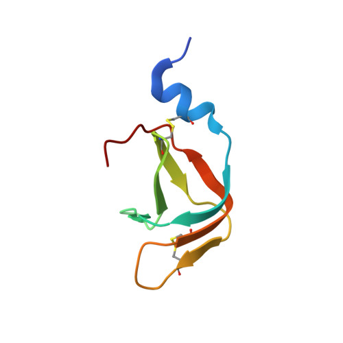
Structural and energetic basis of infection by the filamentous bacteriophage IKe.

Jakob, R.P.Geitner, A.J.Weininger, U.Balbach, J.Dobbek, H.Schmid, F.X.

(2012) Mol Microbiol 84: 1124-1138

  • DOI: https://doi.org/10.1111/j.1365-2958.2012.08079.x
  • Primary Citation of Related Structures:  
    4EO0, 4EO1

  • PubMed Abstract: 

    Filamentous phage use the two N-terminal domains of their gene-3-proteins to initiate infection of Escherichia coli. One domain interacts with a pilus, and then the other domain binds to TolA at the cell surface. In phage fd, these two domains are tightly associated with each other, which renders the phage robust but non-infectious, because the TolA binding site is inaccessible. Activation for infection requires partial unfolding, domain disassembly and prolyl isomerization. Phage IKe infects E. coli less efficiently than phage fd. Unlike in phage fd, the pilus- and TolA-binding domains of phage IKe are independent of each other in stability and folding. The site for TolA binding is thus always accessible, but the affinity is very low. The structures of the two domains, analysed by X-ray crystallography and by NMR spectroscopy, revealed a unique fold for the N-pilus-binding domain and a conserved fold for the TolA-binding domain. The absence of an activation mechanism as in phage fd and the low affinity for TolA probably explain the low infectivity of phage IKe. They also explain why, in a previous co-evolution experiment with a mixture of phage fd and phage IKe, all hybrid phage adopted the superior infection mechanism of phage fd.


  • Organizational Affiliation

    Laboratorium für Biochemie and Bayreuther Zentrum für Molekulare Biowissenschaften, Universität Bayreuth, D-95440 Bayreuth, Germany.


Macromolecules
Find similar proteins by:  (by identity cutoff)  |  3D Structure
Entity ID: 1
MoleculeChains Sequence LengthOrganismDetailsImage
Attachment protein G3P70Salmonella phage IKeMutation(s): 0 
Gene Names: III
UniProt
Find proteins for P03663 (Salmonella phage IKe)
Explore P03663 
Go to UniProtKB:  P03663
Entity Groups  
Sequence Clusters30% Identity50% Identity70% Identity90% Identity95% Identity100% Identity
UniProt GroupP03663
Sequence Annotations
Expand
  • Reference Sequence
Small Molecules
Ligands 1 Unique
IDChains Name / Formula / InChI Key2D Diagram3D Interactions
MG
Query on MG

Download Ideal Coordinates CCD File 
B [auth A]MAGNESIUM ION
Mg
JLVVSXFLKOJNIY-UHFFFAOYSA-N
Experimental Data & Validation

Experimental Data

  • Method: X-RAY DIFFRACTION
  • Resolution: 1.80 Å
  • R-Value Free: 0.219 
  • R-Value Work: 0.187 
  • R-Value Observed: 0.190 
  • Space Group: P 21 21 21
Unit Cell:
Length ( Å )Angle ( ˚ )
a = 28.55α = 90
b = 33.81β = 90
c = 65.59γ = 90
Software Package:
Software NamePurpose
MAR345dtbdata collection
AMoREphasing
BUSTERrefinement
XDSdata reduction
XDSdata scaling

Structure Validation

View Full Validation Report


Entry History 

Deposition Data

Revision History  (Full details and data files)

  • Version 1.0: 2012-05-30
    Type: Initial release
  • Version 1.1: 2013-01-09
    Changes: Database references
  • Version 1.2: 2017-10-25
    Changes: Author supporting evidence
  • Version 1.3: 2023-09-13
    Changes: Data collection, Database references, Derived calculations, Refinement description