


crystal structure of DNA ligase
Wang, T., Xu, W., Charifson, P., Wei, Y.To be published.
Experimental Data Snapshot
Entity ID: 1 | |||||
|---|---|---|---|---|---|
| Molecule | Chains | Sequence Length | Organism | Details | Image |
| DNA ligase | 331 | Enterococcus faecalis | Mutation(s): 0 EC: 6.5.1.2 | 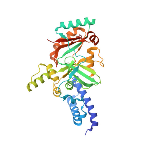 | |
UniProt | |||||
Entity Groups | |||||
| Sequence Clusters | 30% Identity50% Identity70% Identity90% Identity95% Identity100% Identity | ||||
| UniProt Group | Q837V6 | ||||
Sequence AnnotationsExpand | |||||
Reference Sequence | |||||
| Ligands 3 Unique | |||||
|---|---|---|---|---|---|
| ID | Chains | Name / Formula / InChI Key | 2D Diagram | 3D Interactions | |
| NMN Download:Ideal Coordinates CCD File | D [auth A] | BETA-NICOTINAMIDE RIBOSE MONOPHOSPHATE C11 H16 N2 O8 P DAYLJWODMCOQEW-TURQNECASA-O |  | ||
| 0OV Download:Ideal Coordinates CCD File | E [auth A] | [4-amino-2-{[(1S,2R)-2-methylcyclohexyl]oxy}-5-oxopyrido[2,3-d]pyrimidin-8(5H)-yl]acetonitrile C16 H19 N5 O2 MZUFVSMPTFUMGZ-PWSUYJOCSA-N |  | ||
| SO4 Download:Ideal Coordinates CCD File | B [auth A], C [auth A] | SULFATE ION O4 S QAOWNCQODCNURD-UHFFFAOYSA-L |  | ||
| Length ( Å ) | Angle ( ˚ ) |
|---|---|
| a = 90.55 | α = 90 |
| b = 86.53 | β = 101.02 |
| c = 57.03 | γ = 90 |
| Software Name | Purpose |
|---|---|
| CrystalClear | data collection |
| CNX | refinement |
| DENZO | data reduction |
| SCALEPACK | data scaling |


