4E36

Crystal structure of the human Endoplasmic Reticulum Aminopeptidase 2 variant N392K


Experimental Data Snapshot

  • Method: X-RAY DIFFRACTION
  • Resolution: 3.22 Å
  • R-Value Free: 0.261 
  • R-Value Work: 0.209 
  • R-Value Observed: 0.211 

wwPDB Validation   3D Report Full Report


Ligand Structure Quality Assessment 


This is version 2.1 of the entry. See complete history


Literature

A common single nucleotide polymorphism in endoplasmic reticulum aminopeptidase 2 induces a specificity switch that leads to altered antigen processing.

Evnouchidou, I.Birtley, J.Seregin, S.Papakyriakou, A.Zervoudi, E.Samiotaki, M.Panayotou, G.Giastas, P.Petrakis, O.Georgiadis, D.Amalfitano, A.Saridakis, E.Mavridis, I.M.Stratikos, E.

(2012) J Immunol 189: 2383-2392

  • DOI: https://doi.org/10.4049/jimmunol.1200918
  • Primary Citation of Related Structures:  
    4E36

  • PubMed Abstract: 

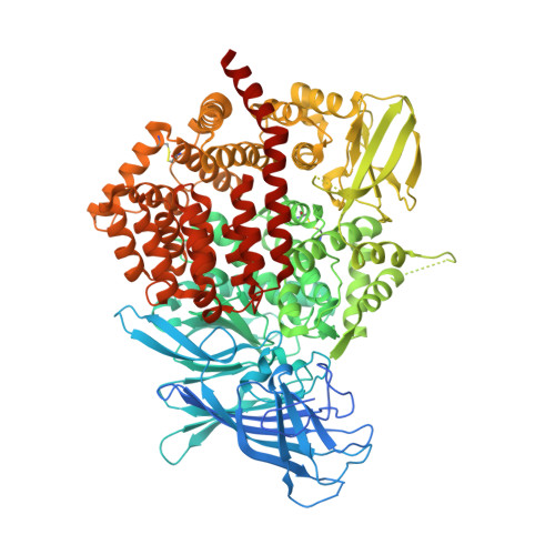
    Endoplasmic reticulum aminopeptidases 1 and 2 (ERAP1 and ERAP2) cooperate to trim antigenic peptide precursors for loading onto MHC class I molecules and help regulate the adaptive immune response. Common coding single nucleotide polymorphisms in ERAP1 and ERAP2 have been linked with predisposition to human diseases ranging from viral and bacterial infections to autoimmunity and cancer. It has been hypothesized that altered Ag processing by these enzymes is a causal link to disease etiology, but the molecular mechanisms are obscure. We report in this article that the common ERAP2 single nucleotide polymorphism rs2549782 that codes for amino acid variation N392K leads to alterations in both the activity and the specificity of the enzyme. Specifically, the 392N allele excises hydrophobic N-terminal residues from epitope precursors up to 165-fold faster compared with the 392K allele, although both alleles are very similar in excising positively charged N-terminal amino acids. These effects are primarily due to changes in the catalytic turnover rate (k(cat)) and not in the affinity for the substrate. X-ray crystallographic analysis of the ERAP2 392K allele suggests that the polymorphism interferes with the stabilization of the N terminus of the peptide both directly and indirectly through interactions with key residues participating in catalysis. This specificity switch allows the 392N allele of ERAP2 to supplement ERAP1 activity for the removal of hydrophobic N-terminal residues. Our results provide mechanistic insight to the association of this ERAP2 polymorphism with disease and support the idea that polymorphic variation in Ag processing enzymes constitutes a component of immune response variability in humans.


  • Organizational Affiliation

    National Center for Scientific Research Demokritos, 15310 Athens, Greece.


Macromolecules
Find similar proteins by:  (by identity cutoff)  |  3D Structure
Entity ID: 1
MoleculeChains Sequence LengthOrganismDetailsImage
Endoplasmic reticulum aminopeptidase 2
A, B
967Homo sapiensMutation(s): 0 
Gene Names: ERAP2IrapLRAP
EC: 3.4.11
UniProt & NIH Common Fund Data Resources
Find proteins for Q6P179 (Homo sapiens)
Explore Q6P179 
Go to UniProtKB:  Q6P179
PHAROS:  Q6P179
GTEx:  ENSG00000164308 
Entity Groups  
Sequence Clusters30% Identity50% Identity70% Identity90% Identity95% Identity100% Identity
UniProt GroupQ6P179
Sequence Annotations
Expand
  • Reference Sequence
Oligosaccharides

Help

Entity ID: 2
MoleculeChains Length2D Diagram Glycosylation3D Interactions
2-acetamido-2-deoxy-beta-D-glucopyranose-(1-4)-2-acetamido-2-deoxy-beta-D-glucopyranose
C, D, F
2N-Glycosylation
Glycosylation Resources
GlyTouCan:  G42666HT
GlyCosmos:  G42666HT
GlyGen:  G42666HT
Entity ID: 3
MoleculeChains Length2D Diagram Glycosylation3D Interactions
alpha-D-mannopyranose-(1-4)-alpha-D-mannopyranose-(1-4)-2-acetamido-2-deoxy-beta-D-glucopyranose-(1-4)-2-acetamido-2-deoxy-beta-D-glucopyranose
E
4N-Glycosylation
Glycosylation Resources
GlyTouCan:  G81874LD
GlyCosmos:  G81874LD
GlyGen:  G81874LD
Small Molecules
Ligands 4 Unique
IDChains Name / Formula / InChI Key2D Diagram3D Interactions
NAG
Query on NAG

Download Ideal Coordinates CCD File 
H [auth A],
I [auth A],
J [auth A],
O [auth B]
2-acetamido-2-deoxy-beta-D-glucopyranose
C8 H15 N O6
OVRNDRQMDRJTHS-FMDGEEDCSA-N
MES
Query on MES

Download Ideal Coordinates CCD File 
K [auth A],
P [auth B]
2-(N-MORPHOLINO)-ETHANESULFONIC ACID
C6 H13 N O4 S
SXGZJKUKBWWHRA-UHFFFAOYSA-N
LYS
Query on LYS

Download Ideal Coordinates CCD File 
G [auth A],
M [auth B]
LYSINE
C6 H15 N2 O2
KDXKERNSBIXSRK-YFKPBYRVSA-O
ZN
Query on ZN

Download Ideal Coordinates CCD File 
L [auth A],
N [auth B]
ZINC ION
Zn
PTFCDOFLOPIGGS-UHFFFAOYSA-N
Experimental Data & Validation

Experimental Data

  • Method: X-RAY DIFFRACTION
  • Resolution: 3.22 Å
  • R-Value Free: 0.261 
  • R-Value Work: 0.209 
  • R-Value Observed: 0.211 
  • Space Group: P 1 21 1
Unit Cell:
Length ( Å )Angle ( ˚ )
a = 74.34α = 90
b = 134.45β = 90.85
c = 127.37γ = 90
Software Package:
Software NamePurpose
MOLREPphasing
PHENIXrefinement
MOSFLMdata reduction
SCALAdata scaling

Structure Validation

View Full Validation Report


Ligand Structure Quality Assessment 


Entry History 

Deposition Data

Revision History  (Full details and data files)

  • Version 1.0: 2012-08-29
    Type: Initial release
  • Version 1.1: 2012-10-17
    Changes: Database references
  • Version 1.2: 2017-11-15
    Changes: Advisory, Refinement description
  • Version 2.0: 2020-07-29
    Type: Remediation
    Reason: Carbohydrate remediation
    Changes: Advisory, Atomic model, Data collection, Database references, Derived calculations, Structure summary
  • Version 2.1: 2023-09-13
    Changes: Advisory, Data collection, Database references, Refinement description, Structure summary