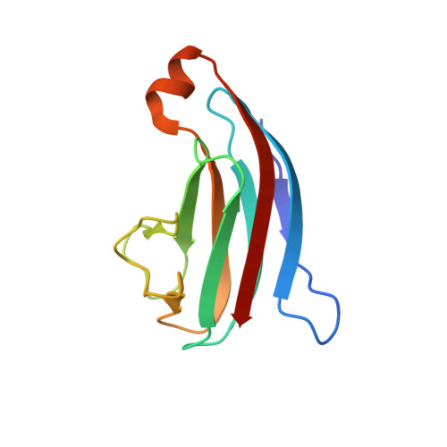
Structural characterization of the PliG lysozyme inhibitor family.
Leysen, S., Vanderkelen, L., Van Asten, K., Vanheuverzwijn, S., Theuwis, V., Michiels, C.W., Strelkov, S.V.(2012) J Struct Biol 180: 235-242
- PubMed: 22634186 Search on PubMed
- DOI: https://doi.org/10.1016/j.jsb.2012.05.006
- Primary Citation Related Structures:
4DY3, 4DY5, 4DZG - PubMed Abstract:
Several Gram-negative bacteria protect themselves against the lytic action of host lysozymes by producing specific proteinaceous inhibitors. So far, four different families of lysozyme inhibitors have been identified including Ivy (Inhibitor of vertebrate lysozyme), MliC/PliC (Membrane associated/periplasmic inhibitor of C-type lysozyme), PliI and PliG (periplasmic inhibitors of I- and G-type lysozymes, respectively). Here we provide the first crystallographic description of the PliG family. Crystal structures were obtained for the PliG homologues from Escherichia coli, Salmonella enterica serotype Typhimurium and Aeromonas hydrophila. These structures show that the fold of the PliG family is very distinct from that of all other families of lysozyme inhibitors. Small-angle X-ray scattering studies reveal that PliG is monomeric in solution as opposed to the dimeric PliC and PliI. The PliG family shares a highly conserved SG(x)xY sequence motif with the MliC/PliC and PliI families where it was shown to reside on a loop that blocks the active site of lysozyme leading to inhibition. Surprisingly, we found that in PliG this motif is not well exposed and not involved in the inhibitory action. Instead, we could identify a distinct cluster of surface residues that are conserved across the PliG family and are essential for efficient G-type lysozyme inhibition, as evidenced by mutagenesis studies.
- Laboratory for Biocrystallography, Department of Pharmaceutical and Pharmacological Sciences, Katholieke Universiteit Leuven, Herestraat 49 bus 822, 3000 Leuven, Belgium.
Organizational Affiliation: