4DQ3 | pdb_00004dq3

Bovine beta-lactoglobulin complex with oleic acid


Experimental Data Snapshot

  • Method: X-RAY DIFFRACTION
  • Resolution: 2.10 Å
  • R-Value Free: 
    0.289 (Depositor), 0.291 (DCC) 
  • R-Value Work: 
    0.231 (Depositor), 0.231 (DCC) 
  • R-Value Observed: 
    0.237 (Depositor) 

Starting Model: experimental
View more details

wwPDB Validation 3D Report Full Report

Validation slider image for 4DQ3

Ligand Structure Quality Assessment 


This is version 1.4 of the entry. See complete history

Literature

Binding of 18-carbon unsaturated fatty acids to bovine beta-lactoglobulin--structural and thermodynamic studies.

Loch, J.I.Bonarek, P.Polit, A.Ries, D.Dziedzicka-Wasylewska, M.Lewinski, K.

(2013) Int J Biol Macromol 57: 226-231

  • DOI: https://doi.org/10.1016/j.ijbiomac.2013.03.021
  • Primary Citation Related Structures: 
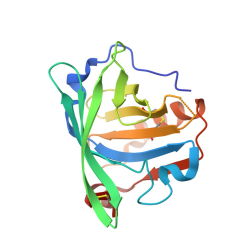
    4DQ3, 4DQ4

  • PubMed Abstract: 

    Binding of 18-carbon unsaturated oleic and linoleic acid to lactoglobulin, the milk protein, has been studied for the first time by isothermal titration calorimetry (ITC) and X-ray crystallography. Crystal structures determined to resolution 2.10 Å have revealed presence of single fatty acid molecule bound in β-barrel, the primary binding site, with carboxyl group hydrogen bonded to Glu62. The aliphatic chain of both ligands is in almost linear conformation and their interactions with the protein are similar to observed in structure of lactoglobulin with stearic acid. The ITC experiments showed that binding of unsaturated fatty acids to LGB is spontaneous and exothermic. The stoichiometry of binding is lower than 1.0, association constant is 9.7 × 10(5)M(-1) and 9.0 × 10(5)M(-1) for oleic and linoleic acid, respectively. Solvent relief seems to be the major contributor to entropic changes upon fatty acid binding to lactoglobulin.


  • Organizational Affiliation
    • Faculty of Chemistry, Jagiellonian University, Ingardena 3, 30-060 Kraków, Poland.

Macromolecule Content 

  • Total Structure Weight: 18.72 kDa 
  • Atom Count: 1,378 
  • Modeled Residue Count: 158 
  • Deposited Residue Count: 162 
  • Unique protein chains: 1

Macromolecules

Find similar proteins by:|  3D Structure
Entity ID: 1
MoleculeChains  Sequence LengthOrganismDetailsImage
Beta-lactoglobulin162Bos taurusMutation(s): 0 
UniProt
Find proteins for P02754 (Bos taurus)
Explore P02754 
Go to UniProtKB:  P02754
Entity Groups
Sequence Clusters30% Identity50% Identity70% Identity90% Identity95% Identity100% Identity
UniProt GroupP02754
Sequence Annotations
Expand
Reference Sequence

Experimental Data & Validation

Experimental Data

  • Method: X-RAY DIFFRACTION
  • Resolution: 2.10 Å
  • R-Value Free:  0.289 (Depositor), 0.291 (DCC) 
  • R-Value Work:  0.231 (Depositor), 0.231 (DCC) 
  • R-Value Observed: 0.237 (Depositor) 
Space Group: P 32 2 1
Unit Cell:
Length ( Å )Angle ( ˚ )
a = 53.46α = 90
b = 53.46β = 90
c = 111.57γ = 120
Software Package:
Software NamePurpose
CrysalisProdata collection
MOLREPphasing
REFMACrefinement
CrysalisProdata reduction
SCALAdata scaling

Structure Validation

View Full Validation Report


Ligand Structure Quality Assessment 


Entry History 

Deposition Data

Revision History  (Full details and data files)

  • Version 1.0: 2012-02-29
    Type: Initial release
  • Version 1.1: 2013-04-03
    Changes: Database references
  • Version 1.2: 2013-08-28
    Changes: Database references
  • Version 1.3: 2023-09-13
    Changes: Data collection, Database references, Derived calculations, Refinement description
  • Version 1.4: 2024-10-16
    Changes: Structure summary