


Target-binding to S100A5 changes Ca(2+)-binding affinity and dynamics.
Liriano, M.A., Varney, K.M., Ishima, R., Toth, E.A., Weber, D.J.To be published.
Experimental Data Snapshot
wwPDB Validation 3D Report Full Report

Entity ID: 1 | |||||
|---|---|---|---|---|---|
| Molecule | Chains | Sequence Length | Organism | Details | Image |
| Protein S100-A5 | 92 | Homo sapiens | Mutation(s): 0 Gene Names: S100A5, S100D | 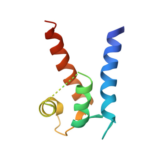 | |
UniProt & NIH Common Fund Data Resources | |||||
PHAROS: P33763 GTEx: ENSG00000196420 | |||||
Entity Groups | |||||
| Sequence Clusters | 30% Identity50% Identity70% Identity90% Identity95% Identity100% Identity | ||||
| UniProt Group | P33763 | ||||
Sequence AnnotationsExpand | |||||
Reference Sequence | |||||
| Ligands 1 Unique | |||||
|---|---|---|---|---|---|
| ID | Chains | Name / Formula / InChI Key | 2D Diagram | 3D Interactions | |
| CA Download:Ideal Coordinates CCD File | C [auth A], D [auth A], E [auth B], F [auth B] | CALCIUM ION Ca BHPQYMZQTOCNFJ-UHFFFAOYSA-N |  | ||
| Length ( Å ) | Angle ( ˚ ) |
|---|---|
| a = 52.692 | α = 90 |
| b = 52.692 | β = 90 |
| c = 67.012 | γ = 90 |
| Software Name | Purpose |
|---|---|
| HKL-2000 | data collection |
| PHASES | phasing |
| REFMAC | refinement |
| HKL-2000 | data reduction |
| HKL-2000 | data scaling |


